Japanese
English
- 有料閲覧
PPV購入案内
1,320円 (1,200円+税10%) こちらは、1120ページから1124ページの文献です。購入には医書.jp本会員登録が必要です。
会員登録完了後に改めて上記「購入サイトへ」を選択してください。
または「購入サイトへ」選択後に「購入ログイン」、「新規会員登録」の順に進んでください。
会員登録について詳しくはをご確認ください。
- Abstract 文献概要
- 1ページ目 Look Inside
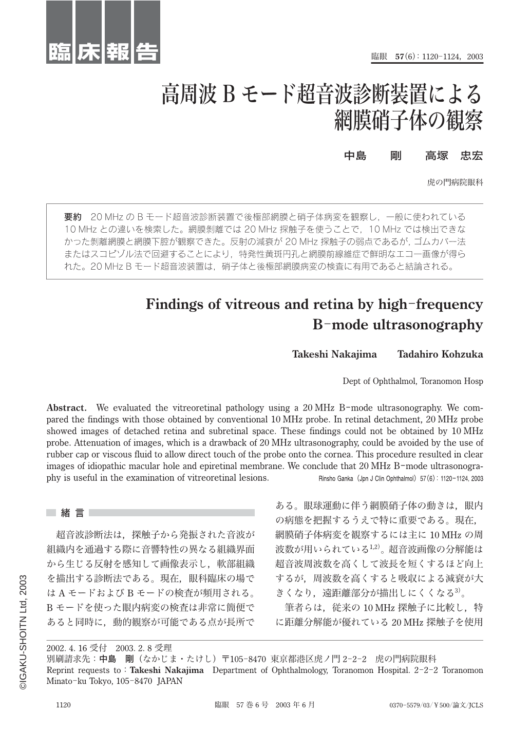
要約 20 MHzのBモード超音波診断装置で後極部網膜と硝子体病変を観察し,一般に使われている10 MHzとの違いを検索した。網膜剝離では20 MHz探触子を使うことで,10 MHzでは検出できなかった剝離網膜と網膜下腔が観察できた。反射の減衰が20 MHz探触子の弱点であるが,ゴムカバー法またはスコピゾル法で回避することにより,特発性黄斑円孔と網膜前線維症で鮮明なエコー画像が得られた。20 MHz Bモード超音波装置は,硝子体と後極部網膜病変の検査に有用であると結論される。
Abstract. We evaluated the vitreoretinal pathology using a 20 MHz B-mode ultrasonography. We compared the findings with those obtained by conventional 10 MHz probe. In retinal detachment,20 MHz probe showed images of detached retina and subretinal space. These findings could not be obtained by 10 MHz probe. Attenuation of images,which is a drawback of 20 MHz ultrasonography,could be avoided by the use of rubber cap or viscous fluid to allow direct touch of the probe onto the cornea. This procedure resulted in clear images of idiopathic macular hole and epiretinal membrane. We conclude that 20 MHz B-mode ultrasonography is useful in the examination of vitreoretinal lesions.
Copyright © 2003, Igaku-Shoin Ltd. All rights reserved.