Japanese
English
- 有料閲覧
PPV購入案内
1,320円 (1,200円+税10%) こちらは、1273ページから1275ページの文献です。購入には医書.jp本会員登録が必要です。
会員登録完了後に改めて上記「購入サイトへ」を選択してください。
または「購入サイトへ」選択後に「購入ログイン」、「新規会員登録」の順に進んでください。
会員登録について詳しくはをご確認ください。
- Abstract 文献概要
- 1ページ目 Look Inside
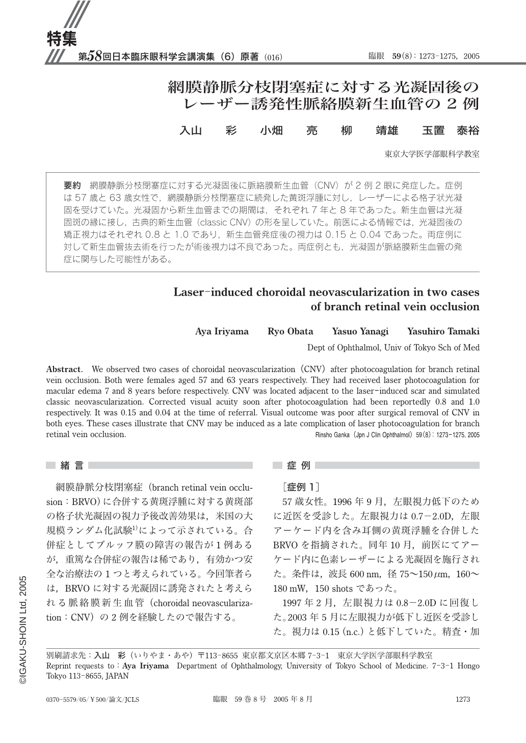
網膜静脈分枝閉塞症に対する光凝固後に脈絡膜新生血管(CNV)が2例2眼に発症した。症例は57歳と63歳女性で,網膜静脈分枝閉塞症に続発した黄斑浮腫に対し,レーザーによる格子状光凝固を受けていた。光凝固から新生血管までの期間は,それぞれ7年と8年であった。新生血管は光凝固斑の縁に接し,古典的新生血管(classic CNV)の形を呈していた。前医による情報では,光凝固後の矯正視力はそれぞれ0.8と1.0であり,新生血管発症後の視力は0.15と0.04であった。両症例に対して新生血管抜去術を行ったが術後視力は不良であった。両症例とも,光凝固が脈絡膜新生血管の発症に関与した可能性がある。
We observed two cases of choroidal neovascularization(CNV)after photocoagulation for branch retinal vein occlusion. Both were females aged 57 and 63 years respectively. They had received laser photocoagulation for macular edema 7 and 8 years before respectively. CNV was located adjacent to the laser-induced scar and simulated classic neovascularization. Corrected visual acuity soon after photocoagulation had been reportedly 0.8 and 1.0 respectively. It was 0.15 and 0.04 at the time of referral. Visual outcome was poor after surgical removal of CNV in both eyes. These cases illustrate that CNV may be induced as a late complication of laser photocoagulation for branch retinal vein occlusion.
Copyright © 2005, Igaku-Shoin Ltd. All rights reserved.