Japanese
English
- 有料閲覧
PPV購入案内
1,320円 (1,200円+税10%) こちらは、95ページから98ページの文献です。購入には医書.jp本会員登録が必要です。
会員登録完了後に改めて上記「購入サイトへ」を選択してください。
または「購入サイトへ」選択後に「購入ログイン」、「新規会員登録」の順に進んでください。
会員登録について詳しくはをご確認ください。
- Abstract 文献概要
- 1ページ目 Look Inside
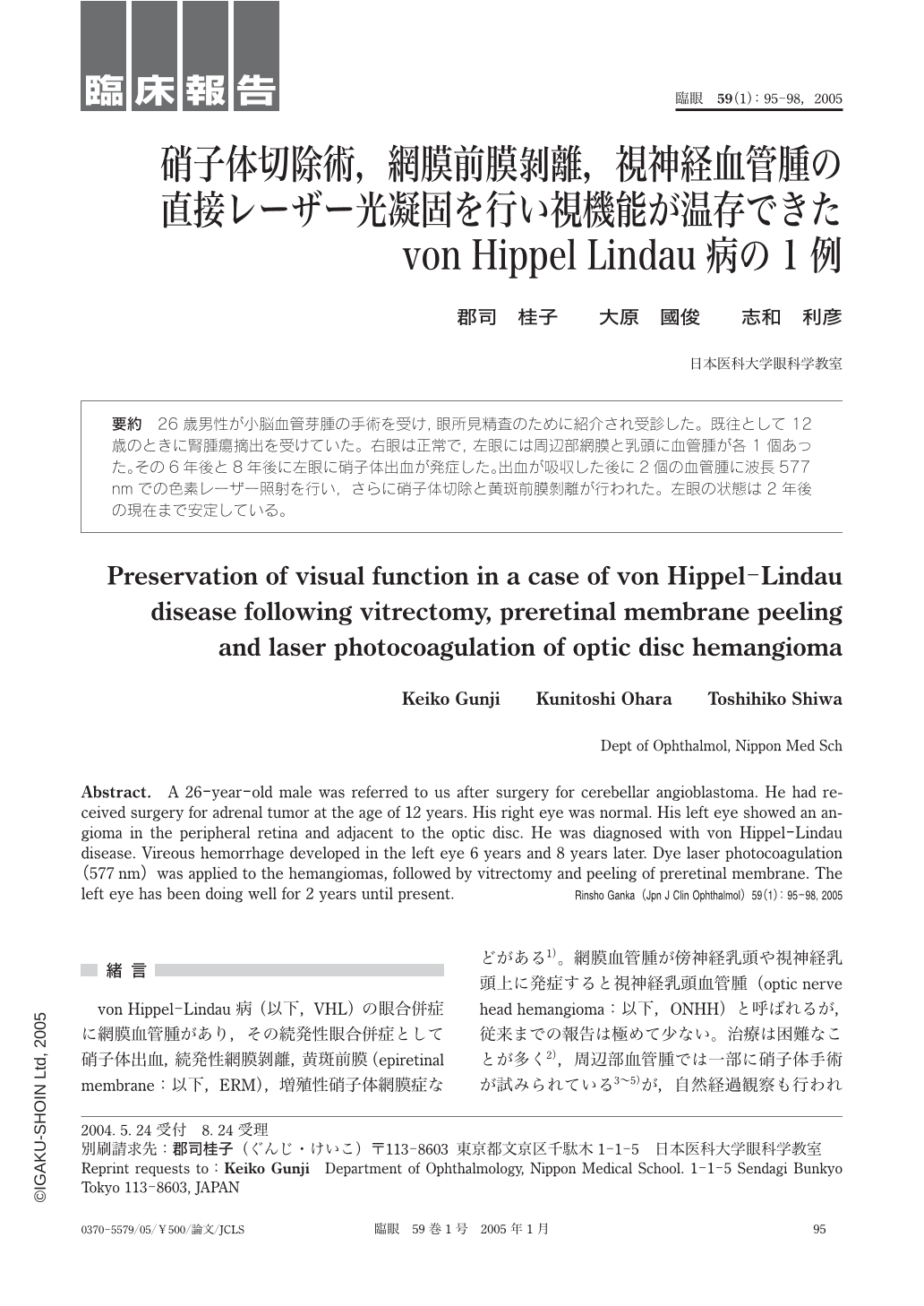
26歳男性が小脳血管芽腫の手術を受け,眼所見精査のために紹介され受診した。既往として12歳のときに腎腫瘍摘出を受けていた。右眼は正常で,左眼には周辺部網膜と乳頭に血管腫が各1個あった。その6年後と8年後に左眼に硝子体出血が発症した。出血が吸収した後に2個の血管腫に波長577nmでの色素レーザー照射を行い,さらに硝子体切除と黄斑前膜剝離が行われた。左眼の状態は2年後の現在まで安定している。
A 26-year-old male was referred to us after surgery for cerebellar angioblastoma. He had received surgery for adrenal tumor at the age of 12 years. His right eye was normal. His left eye showed an angioma in the peripheral retina and adjacent to the optic disc. He was diagnosed with von Hippel-Lindau disease. Vireous hemorrhage developed in the left eye 6 years and 8 years later. Dye laser photocoagulation(577nm)was applied to the hemangiomas,followed by vitrectomy and peeling of preretinal membrane. The left eye has been doing well for 2 years until present.
Copyright © 2005, Igaku-Shoin Ltd. All rights reserved.