Japanese
English
- 有料閲覧
PPV購入案内
1,320円 (1,200円+税10%) こちらは、325ページから329ページの文献です。購入には医書.jp本会員登録が必要です。
会員登録完了後に改めて上記「購入サイトへ」を選択してください。
または「購入サイトへ」選択後に「購入ログイン」、「新規会員登録」の順に進んでください。
会員登録について詳しくはをご確認ください。
- Abstract 文献概要
- 1ページ目 Look Inside
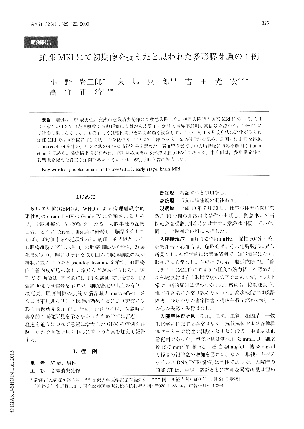
症例は,57歳男性。突然の意識消失発作にて救急入院した。初回入院時の頭部MRIにおいて,T1は正常だがT2では左側頭葉から頭頂葉に皮質から皮質下にかけて境界不鮮明な高信号を認めた。Gd-T1にて造影効果はなかった。腫瘍もしくは変性疾患を考え経過を観察していたが,約4カ月後症状の悪化がみられ頭部MRIでは同部位にT1で明らかな低信号,T2にて内部が不均一な高信号域を認め,周囲には広範な浮腫とmass effectを伴い,リング状の不整な造影効果を認めた。脳血管撮影では中大脳動脈に境界不鮮明なtumorstainを認めた。腫瘍摘出術が行われ、病理組織検査は多形膠芽腫(GBM)であった。本症例は,多形膠芽腫の初期像を捉えた貴重な症例であると考えられ,鑑別診断を含め報告した。
A 57-year-old male was urgently carried to our hos-pital because of sudden loss of consciousness, lasting about 10 minutes. He had resumed consciousness be-fore he arrived at our hospital. Neurologically, he had mild muscle weakness of the right arm. Deep tendon reflexes in the right upper extremity were reduced. In high level functions, speech disturbance, dysgraphia (disturbed ability to write Hiragana), and constructive apraxia were noted. A brain MRI upon admission showed a poorly demarcated , high signal intensity area in the cortical and subcortical layers of the left temporal and parietal lobes.
Copyright © 2000, Igaku-Shoin Ltd. All rights reserved.