Japanese
English
- 有料閲覧
PPV購入案内
1,320円 (1,200円+税10%) こちらは、383ページから385ページの文献です。購入には医書.jp本会員登録が必要です。
会員登録完了後に改めて上記「購入サイトへ」を選択してください。
または「購入サイトへ」選択後に「購入ログイン」、「新規会員登録」の順に進んでください。
会員登録について詳しくはをご確認ください。
- Abstract 文献概要
- 1ページ目 Look Inside
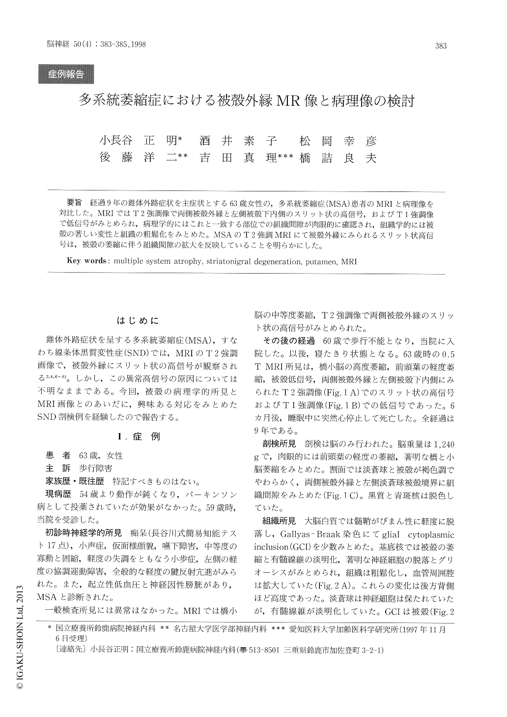
経過9年の錐体外路症状を主症状とする63歳女性の,多系統萎縮症(MSA)患者のMRIと病理像を対比した。MRIではT2強調像で両側被殻外縁と左側被殻下内側のスリット状の高信号,およびT1強調像で低信号がみとめられ,病理学的にはこれと一致する部位での組織間隙が肉眼的に確認され,組織学的には被殻の著しい変性と組織の粗鬆化をみとめた。MSAのT2強調MRIにて被殻外縁にみられるスリット状高信号は,被殻の萎縮に伴う組織間隙の拡大を反映していることを明らかにした。
The slit hyperintensity of the lateral margin of the putamen in T2 weighted MRI is a characteristic finding in those patients with multiple system atro-phy (MSA) involving extrapyramidal system. In spite of some speculations such as demyelination, gliosis, iron deposition or increased extracellular fluid, the nature of the abnormal signal intensity has still been remained uncertain.
In this paper, we report the coincidental findings of pathology and magnetic resonance imaging of the putaminal margin in a case of MSA.
Copyright © 1998, Igaku-Shoin Ltd. All rights reserved.