Japanese
English
- 有料閲覧
PPV購入案内
1,320円 (1,200円+税10%) こちらは、1191ページから1195ページの文献です。購入には医書.jp本会員登録が必要です。
会員登録完了後に改めて上記「購入サイトへ」を選択してください。
または「購入サイトへ」選択後に「購入ログイン」、「新規会員登録」の順に進んでください。
会員登録について詳しくはをご確認ください。
- Abstract 文献概要
- 1ページ目 Look Inside
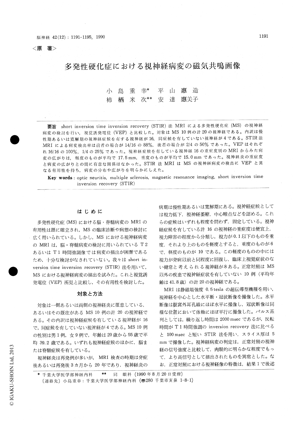
short inversion time inversion recovery(STIR)法MRIによる多発性硬化症(MS)の視神経病変の検討を行い,視覚誘発電位(VEP)と比較した。対象はMS 10例の計20の視神経である。内訳は慢性期あるいは寛解期の視神経症候を有する視神経が16,同症候を有していない視神経が4である。STIR法MRIによる病変検出率は前者の場合が14/16の88%,後者の場合が2/4の50%であった。VEPはそれぞれ16/16の100%,1/4の25%であった。視神経症候を有している視神経16の重症度別のMRIからみた病変の広がりは,軽度のものが平均で17.5mm,重度のものが平均で15.0mmであった。視神経炎の重症度と病変の広がりとの間に有意な関係はなかった。STIR法MRIはMSの視神経病変の検出にVEPと異なる有用性を持ち,病変の分布や広がりを明らかにしえた。
Magnetic resonance imaging (MRI) of the optic nerves was performed in 10 patients with multiple sclerosis (MS) using short inversion time inversion recovery (STIR) pulse sequences, and the results were compared with the visual evoked potentials (VEP).
The 10 patients had optic neuritis in the chronic or remitting phase together with additional symp-toms or signs allowing a diagnosis of clinically definite or probable MS. Sixteen optic nerves were clinically affected and 4 were unaffected. MRI was performed using a 0.5 tesla supercon-ducting unit, and multiple continuous 5 mm coronal and axial STIR images were obtained. A lesion was judged to be present if a focal or diffuse area of increased signal intensity was detected in the optic nerve. In VEP, a delay in peak latency or no P 100 component was judged to be abnormal.
With regard to the clinically affected optic ner-ves, MRI revealed a region of increased signal in-tensity in 14/16 (88%) and the VEP was abnormal in 16/16 (100%). In the clinically unaffected optic nerves, MRI revealed an increased signal intensity in 2/4 (50%). One of these nerves had an abnor-mal VEP and the other had a VEP latency at the upper limit of normal. The VEP was abnormal in 1/4 (25%). In the clinically affected optic nerves, the degree of loss of visual acuity was not associ-ated with the longitudinal extent of the lesions shown by MRI. The mean length was 17.5 mm in optic nerves with a slight disturbance of visual acuity and 15.0 mm in nerves with severe visual loss.
MRI using STIR pulse sequences was found to be almost as sensitive as VEP in detecting both clini-cally affected and unaffected optic nerve lesions in patients with MS, and was useful in visualizing the location or size of the lesions.
Copyright © 1990, Igaku-Shoin Ltd. All rights reserved.