Japanese
English
- 有料閲覧
- Abstract 文献概要
- 1ページ目 Look Inside
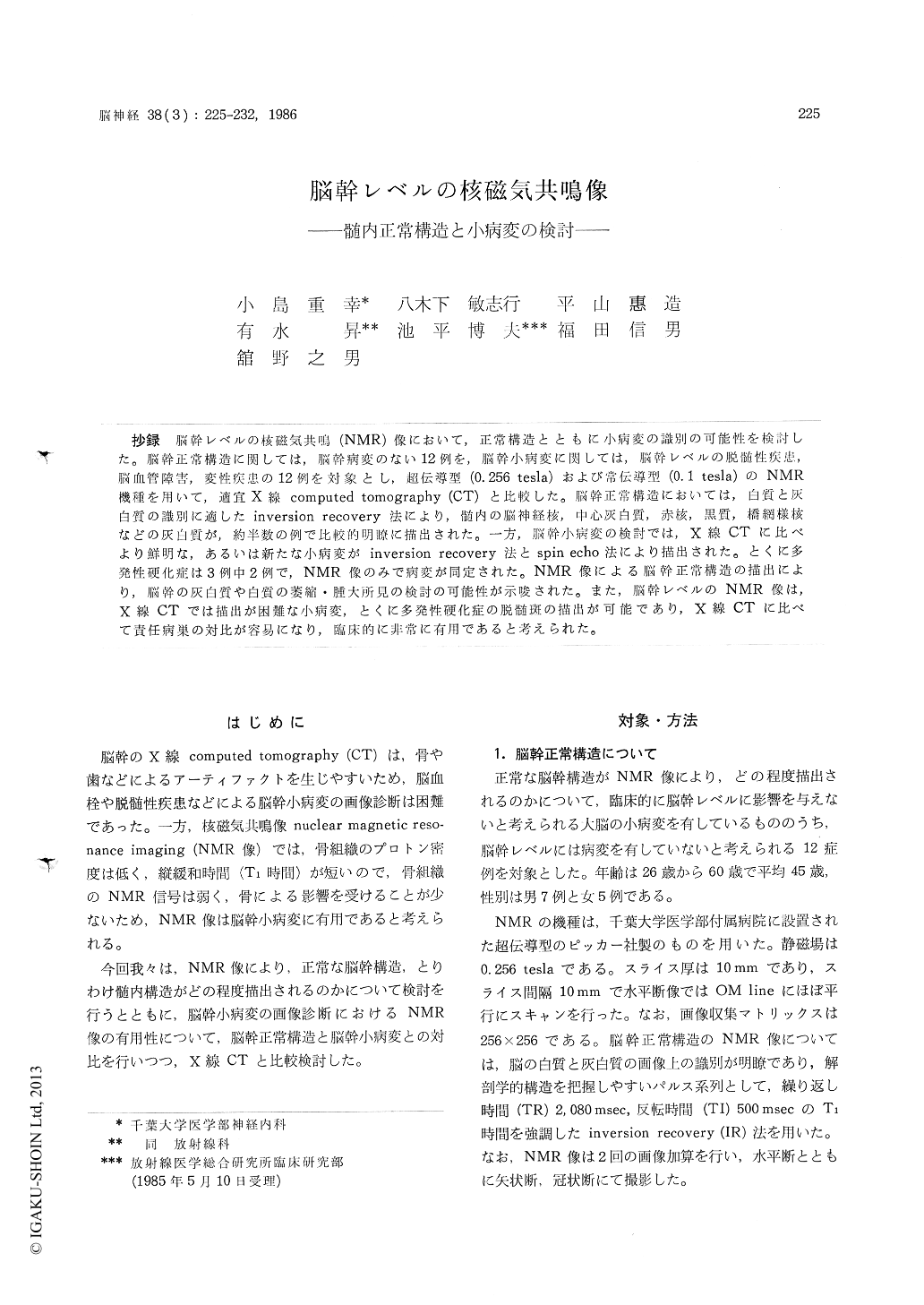
抄録 脳幹レベルの核磁気共鳴(NMR)像において,正常構造とともに小病変の識別の可能性を検討した。脳幹正常構造に関しては,脳幹病変のない12例を,脳幹小病変に関しては,脳幹レベルの脱髄性疾患,脳血管障害,変性疾患の12例を対象とし,超伝導型(0.256tesla)および常伝導型(0.1tesla)のNMR機種を用いて,適宜X線computed tomography (CT)と比較した。脳幹正常構造においては,白質と灰白質の識別に適したinversion recovery法により,髄内の脳神経核,中心灰白質,赤核,黒質,橋網様核などの灰白質が,約半数の例で比較的明瞭に描出された。一方,脳幹小病変の検討では,X線CTに比べより鮮明な,あるいは新たな小病変がinversion recovery法とspin echo法により描出された。とくに多発性硬化症は3例中2例で,NMR像のみで病変が同定された。 NMR像による脳幹正常構造の描出により,脳幹の灰白質や白質の萎縮・腫大所見の検討の可能性が示唆された。また,脳幹レベルのNMR像は,X線CTでは描出が困難な小病変,とくに多発性硬化症の脱髄斑の描出が可能であり, X線CTに比べて責任病巣の対比が容易になり,臨床的に非常に有用であると考えられた。
Nuclear magntic resonance (NMR) imaging of the brainstem region from 12 asymptomatic indivi-duals were reviewed in addition to these of 12 patients with various symptoms of small brain-stem lesions. Abnormalities consisted of 3 cases of multiple sclerosis, 1 case of neuro-Behget disease, 5 cases of infarction and hematoma and 3 cases of degenerative disease.
NMR transverse imaging using inversion reco-very sequence was able to locate many of the normal intra-axial brainstem nuclei, such as the periaqueductal gray matter, the red nucleus, the substantia nigra, the pontine nuclei, the pontine reticular nuclei, the facial nerve nucleus and soon in an about half of 12 asymptomatic individu-als. The remarkable gray-white matter differentia-tion was obtained on NMR imaging using inver-sion recovery sequence and enabled the internal structures to be visualized within the brainstem. In addition, the midsagittal imaging provided an excellent demonstration of anatomical relation-ships of the brainstem and surrounding structures. In the diencephalic region, the mamillary body, the anterior commissure and the optic chiasma were also demonstrated on the midsagittal ima-ging.
The lesions within the brainstem were vaguely shown on X-ray computed tomography in 6 of 12 patients but NMR imaging using inversion reco-very or spin echo sequence provided more de-tailed data and revealed clear small lesions, such as the demyelinated plaques of multiple sclerosis and lacunar infarcts in 9 of 12 patients. Espe-cially, in 2 of 3 multiple sclerosis patients, the plaques of the brainstem were definitely iden-tified on NMR imaging only and the accurate lo-calized lesion which was responsible for the facial myokymia or the Foville syndrome was identified. These studies results show that on NMR ima-ging using several pulse sequences, it is possible to examine the atrophic or hypertrophic findings of the brainstem internal structures and compare the localization of the lesions with clinical symp-toms accurately.

Copyright © 1986, Igaku-Shoin Ltd. All rights reserved.


