Japanese
English
- 有料閲覧
PPV購入案内
1,320円 (1,200円+税10%) こちらは、583ページから588ページの文献です。購入には医書.jp本会員登録が必要です。
会員登録完了後に改めて上記「購入サイトへ」を選択してください。
または「購入サイトへ」選択後に「購入ログイン」、「新規会員登録」の順に進んでください。
会員登録について詳しくはをご確認ください。
- Abstract 文献概要
- 1ページ目 Look Inside
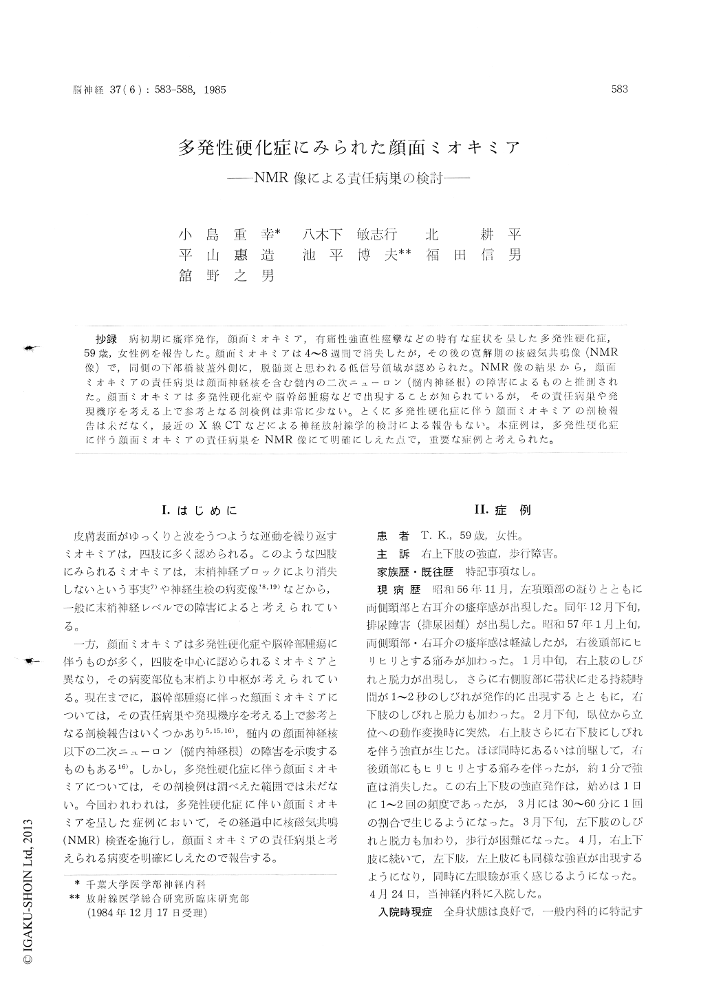
抄録 病初期に掻痒発作,顔面ミオキミア,有痛性強直性痙攣などの特有な症状を呈した多発性硬化症,59歳,女性例を報告した。顔面ミオキミアは4〜8週間で消失したが,その後の寛解期の核磁気共鳴像(NMR像)で,同側の下部橋被蓋外側に,脱髄斑と思われる低信号領域が認められた。 NMR像の結果から,顔面ミオキミアの責任病巣は顔面神経核を含む髄内の二次ニューロン(髄内神経根)の障害によるものと推測された。顔面ミオキミアは多発性硬化症や脳幹部腫瘍などで出現することが知られているが,その責任病巣や発現機序を考える上で参考となる剖検例は非常に少ない。とくに多発性硬化症に伴う顔面ミオキミアの剖検報告は未だなく,最近のX線CTなどによる神経放射線学的検討による報告もない。本症例は,多発性硬化症に伴う顔面ミオキミアの責任病巣をNMR像にて明確にしえた点で,重要な症例と考えられた。
A 59-year-old female of facial myokymia with multiple sclerosis was reported. In this case, facial myokymia appeared at the same time as the first attack of multiple sclerosis, in association with paroxysmal pain and desesthesia of the neck, painful tonic seizures of the right upper and lower extremities and cervical transverse myelo-pathy. The facial myokymia consisted of grossly visible, continuous, fine and worm-like movement, which often began in the area of the left orbicu-laris oculi and spread to the other facial muscles on one side.
Electromyographic studies revealed grouping of motor units and continuous spontaneous rhyth-mic discharges in the left orbicularis oris sug-gesting facial myokymia, but there were no ab-normalities on voluntary contraction. Sometimesdoublet or multiplet patterns occurred while at other times the bursts were of single motor po-tential. The respective frequencies were 3-4/sec and 40-50/sec. There was no evidence of fibrillation. The facial myokymia disappeared after 4-8 weeks of administration of prednisolone and did not recur.
In the remission stage after disappearance of the facial myokymia, nuclear magnetic resonance (NMR) imaging by the inversion recovery method demonstrated low intensity demyelinated plaque in the left lateral tegmentum of the inferior pons, which was responsible for the facial myokymia, but X-ray computed tomography revealed no pa-thological findings. The demyelinated plaque de-monstrated by NMR imaging seemed to be located in the infranuclear area of the facial nerve nucleus and to involve the intramedurally root.
It is known that facial myokymia is found especially in association with multiple sclerosis and brain stem tumors, and the location of the lesion causing facial myokmia is thought to be within the brain stem. But it is uncertain whether the lesion is a supra-, para- or infranuclear area of the facial nerve nucleus, since there are few clinicopathological studies on facial myokymia with brain stem tumors and no studies on multiple sclerosis.
The present case was reported because of the rarity of previous reports, and because NMR imaging demonstrated a lesion surmised to be the cause of the facial myokymia with multiple sclerosis.
Copyright © 1985, Igaku-Shoin Ltd. All rights reserved.