Japanese
English
- 有料閲覧
PPV購入案内
1,320円 (1,200円+税10%) こちらは、187ページから193ページの文献です。購入には医書.jp本会員登録が必要です。
会員登録完了後に改めて上記「購入サイトへ」を選択してください。
または「購入サイトへ」選択後に「購入ログイン」、「新規会員登録」の順に進んでください。
会員登録について詳しくはをご確認ください。
- Abstract 文献概要
- 1ページ目 Look Inside
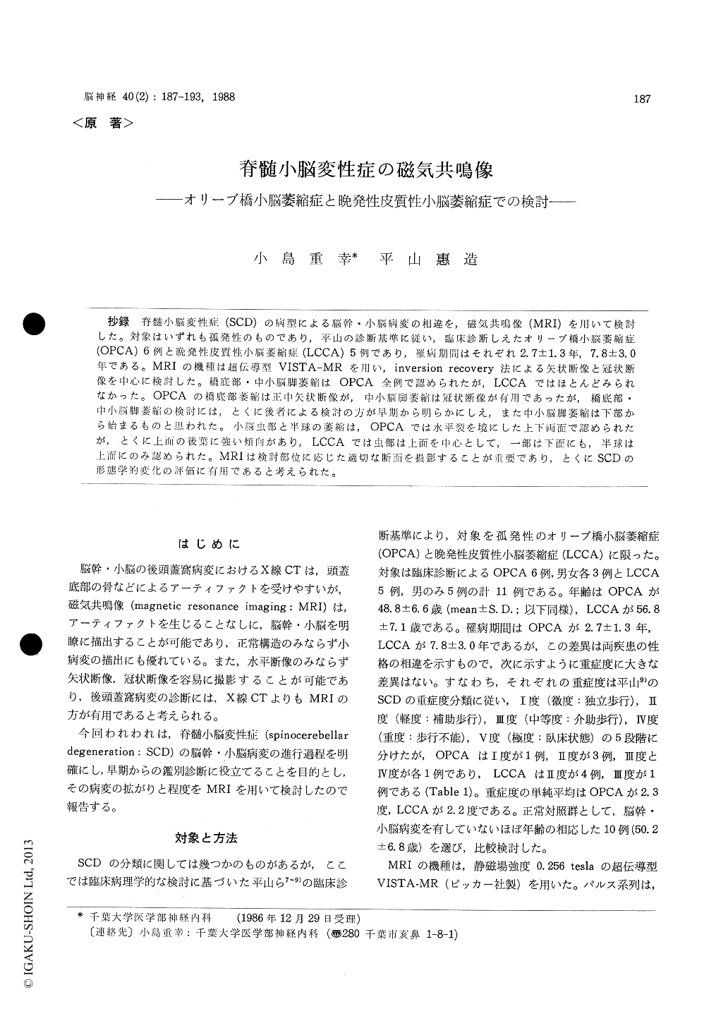
抄録 脊髄小脳変性症(SCD)の病型による脳幹・小脳病変の相違を,磁気共鳴像(MRI)を用いて検討した。対象はいずれも孤発性のものであり,平山の診断基準に従い,臨床診断しえたオリーブ橋小脳萎縮症(OPCA)6例と晩発性皮質性小脳萎縮症(LCCA)5例であり,罹病期間はそれぞれ2.7±1.3年,7.8±3.0年である。MRIの機種は超伝導型VISTA-MRを用い,inversion recovery法による矢状断像と冠状断像を中心に検討した。橋底部・中小脳脚萎縮はOPCA全例で認められたが,LCCAではほとんどみられなかった。OPCAの橋底部萎縮は正中矢状断像が,中小脳脚萎縮は冠状断像が有用であったが,橋底部・中小脳脚萎縮の検討には,とくに後者による検討の方が早期から明らかにしえ,また中小脳脚萎縮は下部から始まるものと思われた。小脳虫部と半球の萎縮は,OPCAでは水平裂を境にした上下両面で認められたが,とくに上面の後葉に強い傾向があり,LCCAでは虫部は上面を中心として,一部は下面にも,半球は上面にのみ認められた。MRIは検討部位に応じた適切な断面を撮影することが重要であり,とくにSCDの形態学的変化の評価に有用であると考えられた。
Magnetic resonance imaging (MRI) was evalu-ated in 11 patients with non-familial spinocerebel-lar degeneration (6 olivo-ponto-cerebellar atrophy (OPCA) and 5 late cortical cerebellar atrophy (LCCA)). MRI was carried out using a super-conducting magnet of 0.256 tesla (VISTA-MR) and an inversion recovery pulse sequence of re-petition time 2.08 sec and inversion time 0.5 sec. The degree of atrophy was assessed with re-gard to ponto-cerebellar system (basis pontis and middle cerebellar peduncle) and cerebellum in the sagittal and coronal images. In the mid-sagittal images, the width of ventral pons, dorsal pons, tegmentum and tectum of midbrain, and the height of fourth ventricle were measured. Especially, the degrees of atrophy of basis pontis in the mid-sagittal image and middle cerebellar peduncle in the coronal image were divided into 4 grades and evaluated respectively. On the other hand, atro-phy of cerebellum was judged from enlargement of cerebellar fissures and reduction of cerebellar volume in the sagittal and coronal images.
Atrophy of ponto-cerebellar system was found in OPCA, but not in LCCA. In OPCA, atrophy of middle cerebellar peduncle in the coronal image, which was likely to begin in an inferior part of pons, was more marked than, or equal to, atrophy of basis pontis in the mid-sagittal image.
With regard to cerebellar vermis, the superior faces were more atrophic than the inferior faces in both OPCA and LCCA but in OPCA, atrophy on the superior faces was dominant in the pos-terior lobe including declive and folium as against dominance in the anterior lobe in LCCA. General-ly, atrophy of cerebellar vermis seemed to bemore marked in OPCA than LCCA compared with the same serious patients. And in OPCA, hemispheric atrophy was also more marked on the superior faces including simple loble and su-perior semilunar loble than the inferior faces. In LCCA, hemispheric atrophy was minimal on the superior faces and was not found on the inferior faces.
MRI seems to be superior to conventional neuro-radiological methods to evaluate the morpholo-gical and degenerative changes in spino-cerebellar degeneration and useful to differentiate OPCA from LCCA early, but in using MRI, it is im-portant to choose the appropriate imaging section and pulse sequences according to the examinating pathological sites and changes.
Copyright © 1988, Igaku-Shoin Ltd. All rights reserved.