Japanese
English
- 有料閲覧
- Abstract 文献概要
- 1ページ目 Look Inside
I.はじめに
孤発性オリーブ橋小脳萎縮症(olivopontocerebellar atrophy of sporadic type;sOPCA)は,神経病理学的には下オリーブ核―橋核―小脳系変性を中核病変とし,これに線条体黒質変性や自律神経病変などを伴う疾患として知られている。sOPCAの小脳歯状核は通常障害されないか,障害されても軽度であるとの記載が多い1,9~11,14)。ところで,sOPCAで歯状核が一般に無傷であるといわれてきたのは,肉眼的,光顕的観察により格別目立つ変化を認めえなかったからであろう。すなわち,病変の有無を神経細胞の脱落の程度や膠細胞の反応の程度から定性的に判断することに頼り,灰白帯そのものの質的,またとくに量的変化についてはほとんど検討がなされてこなかった。しかし,近年は画像解析装置の導入によって,定量的解析にもとづく病変の詳細な検討が可能となってきた。筆者らは,画像解析装置を用いて正常対照の歯状核の形態を明らかにするとともに,遺伝性運動失調症を中心に歯状核の障害様式についてこれまで検討を行なってきた4~8)。
今回は,sOPCAの歯状核の障害の性格と特徴について,神経細胞数,神経細胞密度,灰白帯の体積などを指標として定量的検討を行なったので報告する。
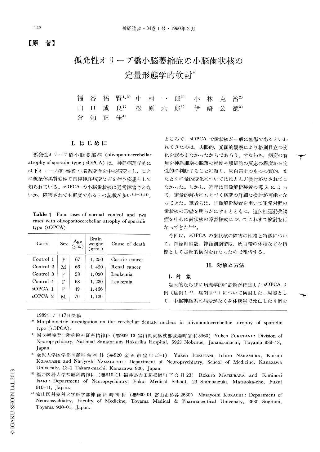
Morphometric investigation was performed on the cerebellar dentate nucleus in two patients with olivopontocerebellar atrophy of sporadic type (sOPCA) and in four normal subjects using computerized image analyzing system. Evaluation of size and population of neurons, neuronal cell density, and total volume of the gray band of the dentate nucleus were examined, after making horizontal serial 20 um thick-sections of the whole cerebellum embedded in celloidin.
In sOPCA, the gray band of the dentate nucleus was reduced so less than 50% in volume of control. Increase of the neuronal cell density was found to be by 170~180%.

Copyright © 1990, Igaku-Shoin Ltd. All rights reserved.


