Japanese
English
- 有料閲覧
- Abstract 文献概要
- 1ページ目 Look Inside
緒言
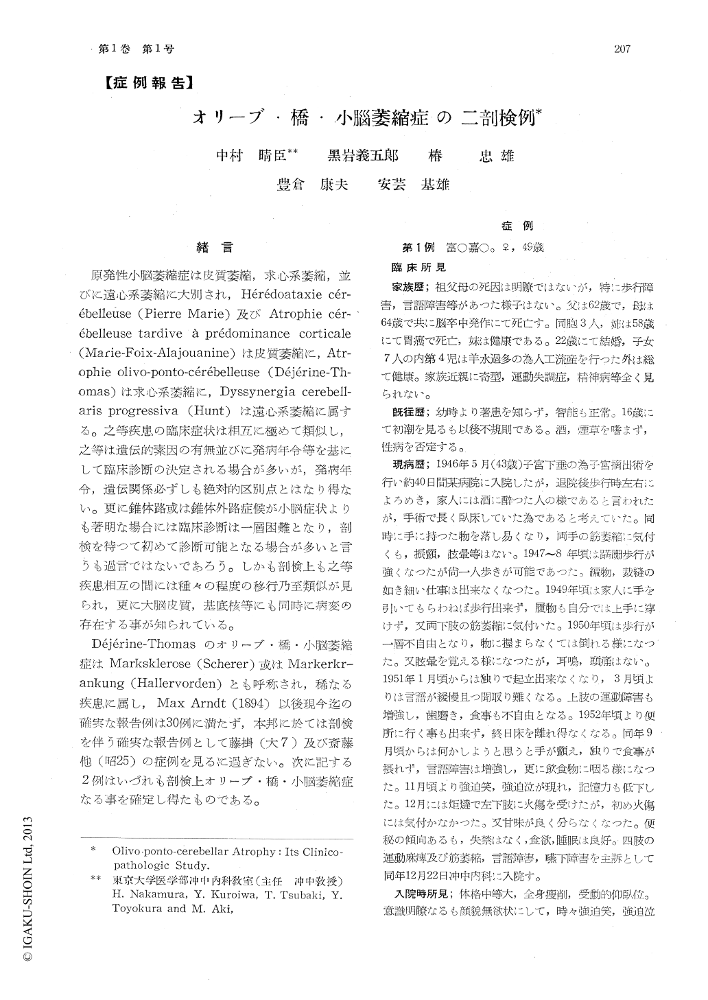
原発性小脳萎縮症は皮質萎縮,求心系萎縮,並びに遠心系萎縮に大別され,Hérédoataxie cérébelleuse(Pierre Marie)及びAtrophie cérébelleuse tardive à prédominance corticale(Marie-Foix-Alajouanine)は皮質萎縮に,Atrophie olivo-ponto-cérébelleuse(Déjérine-Thomas)は求心系萎縮に,Dyssynergia cerebellaris Progressiva(Hunt)は遠心系萎縮に属する。之等疾患の臨床症状は相互に極めて類似し,之等は遺伝的素因の有無並びに発病年令等を基にして臨床診断の決定される場合が多いが,発病年令,遺伝関係必ずしも絶対的区別点とはなり得ない。更に錐体路或は錐体外路症候が小脳症状よりも著明な場合には臨床診断は一層困難となり,剖検を待つて初めて診断可能となる場合が多いと言うも過言ではないであろう。しかも剖検上も之等疾患相互の間には種々の程度の移行乃至類似が見られ,更に大脳皮質,基底核等にも同時に病変の存在する事が知られている。
Case I: K.T., a housewife, aged 49, was not hereditarily tainted. After hysterectomy at age 43, she noted that her gait became ataxic, which was followed by dizziness and disturbance of speech. These symptoms got worse gradually until she developed intention tremor, spastic tetraplegia, muscular atrophy in the distal parts of the extremities and disturbance in swallowing. Emotional incontinence, lowering of the mental activity and finally bladder and rectal disturbances appeared. She died about 6 years after the onset of the illnes.
Postmortem examination revealed that the brain weighed 1040gr., looked atrophic generally. Atrophy of the cerebellum and flattening of the basilar portion of the pons were particularly marked. Microscopically, there were reduction and degenerative changes of the ganglion cells in the pontile nuclei, reticlo-tegmental nucleus, olivary nuclei and arcuate nucleus, and increase of the glia cells in these nuclei. Marked demyelination and gliosis were also observed in the transverse fibers of the pons, the olivocerebellar fibers and the central white matter of the cerebellum. No pathological changes were found in the cerebellar cortex, except the mild reduction of thePurkinje cells. The dentate nucleus and the superior cerebellar peduncle were intact. Demyelination and gliosis in the pyramid and in the lateral corticospinal tract, reduction and degenerative changes of the anterior horn cells, and mild gliosis of the posterior funiculus were observed. No definite pathologic change was found in the lateral cuneate nucleus and in the lateral reticular nucleus. Scattering of melanin pigments was seen outside the nerve cells in the substantia nigra, but some of that pigments were included in the glia cells while others were outside them.
Case II: K. K., a nurseryman, aged 73, had nothing to be mentioned in his family history. He had a mild cerebral attack, when he was 68 years old, which was followed by paraparesis. Disturbance of speech, tremor and ataxia were gradually manifested and muscular rigidity developed during the course. Mild hypesthesia and positive Rossolimo and Mendel-Bechterew's signs were noted on the left leg, and finally bladder and rectal disturbances appeared. He died about 5 years after the onset of the illness.
The brain weighed 1176gr. Microscopically, atrophy of the cerebellum, the basilar portion of the pons and the olive was found. An old softening area in the left insula and mild atherosclerosis in the basilar artery were seen. Microscopically, almost the same changes as in the first case were observed in the cerebellum, pontile nuclei, reticlotegmental nucleus, olvar nuclei, arcuate nucleus, transverse fibers of the pons, olivocerebellar fibers and substantia nigra. Gliosis was remarkable in the caudate nucleus and the putamen. The spinal cord was not studied.
Chief lesions in these two cases were degenerative changes in the system of the corpus pontobulbare (Esseck) and therefore, it is concluded that these are atrophie olivo-ponto-cerebelleuse (Dejerine-Thomas). However, the first case was complicated with the pathological changes of amyotrophic lateral sclerosis, while the second case was found to have gliosis in the corpus striatum. Clinical and pathological observations of these two cases were compared with that of hitherto reported literatures.

Copyright © 1956, Igaku-Shoin Ltd. All rights reserved.


