Japanese
English
- 有料閲覧
PPV購入案内
1,320円 (1,200円+税10%) こちらは、621ページから626ページの文献です。購入には医書.jp本会員登録が必要です。
会員登録完了後に改めて上記「購入サイトへ」を選択してください。
または「購入サイトへ」選択後に「購入ログイン」、「新規会員登録」の順に進んでください。
会員登録について詳しくはをご確認ください。
- Abstract 文献概要
- 1ページ目 Look Inside
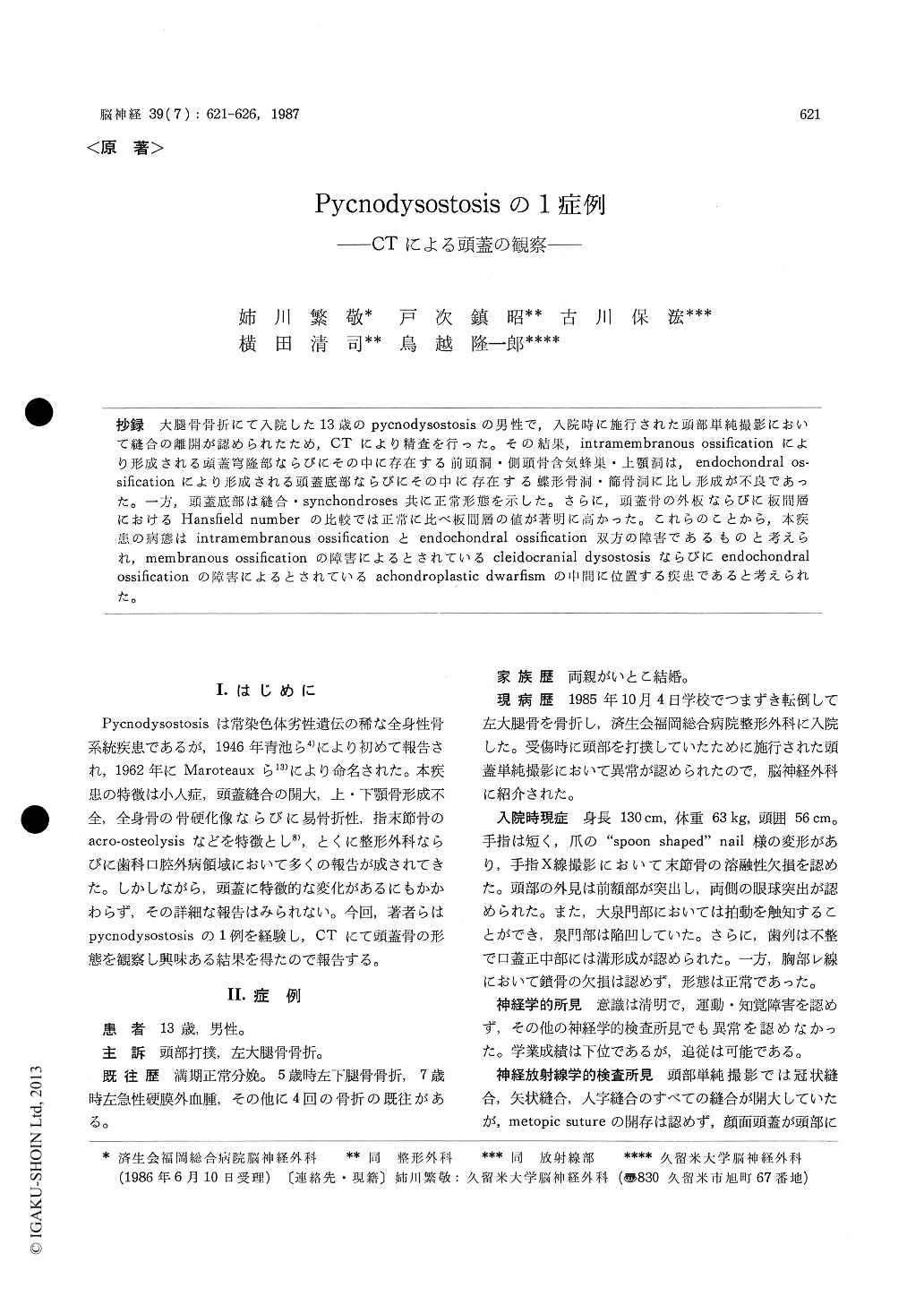
抄録 大腿骨骨折にて入院した13歳のpycnodysostosisの男性で,入院時に施行された頭部単純撮影において縫合の離開が認められたため,CTにより精査を行った。その結果,intramembranous ossificationにより形成される頭蓋穹隆部ならびにその中に存在する前頭洞・側頭骨含気蜂巣・上顎洞は,endochondral os—sificationにより形成される頭蓋底部ならびにその中に存在する蝶形骨洞・篩骨洞に比し形成が不良であった。一方,頭蓋底部は縫合・synchondroses共に正常形態を示した。さらに,頭蓋骨の外板ならびに板間層におけるHansfield numberの比較では正常に比べ板間層の値が著明に高かった。これらのことから,本疾患の病態はintramembranous ossificationとendochondral ossification双方の障害であるものと考えられ’membranous ossificationの障害によるとされているcleidocranial dysostosisならびにendochondralossificationの障害によるとされているachondroplastic dwarfismの中間に位置する疾患であると考えられた。
A 13-year-old boy was presented to the De-partment of Neurosurgery, Saiseikai Fukuoka Gene-ral Hospital for further examinations concerning abnormal findings in the skull radiogram taken when he struck his head.
His physical features showed some characteris-tics the same as those of pycnodysostosis as fol-lows-proportionate dwarfism, prominent forehead, short spoon-shaped fingers, bilateral exophthalmos.
A skull radiogram revealed widely open cra-nial sutures with no healing of the fracture and craniotomy which was performed for an acute epidural hematoma 6 years ago. Furthermore, the mandible was hypoplastic with a virtural loss of mandibular angle.
CT of the soft tissues showed somewhat dilatedcortical sulci and ventricles without any structural abnormalities in the brain. CT of bone algo-rythum revealed specific characteristics of this dis-ease. The paranasal sinuses were quite hypoplastic. Especially in the maxillary sinuses, frontal sinus-sus and mastoid air cells, none of developments of sinuses were noted, even though the middle and internal ear seemed to be normal. Moreover, the ethomoid and sphenoid sinuses were noted, although their developments were poor. The ap-pearance of skull base was normal, including the inlets and outlets of cranial nerves or vessels and synchondroses. However, the density of the skull base, especially in the diploe, was higher than normal in Hansfield number. Furthermore, detailed measurements of skull base demonstrated that the skull base itself was also dwarfish.
Pycnodysostosis is a generalized skeletal disease whose cardinal features are moderate generalized osteosclerosis and dwarfism. However, the detail-led observation on the cranium by CT has not been reported. In our study, the development of sinuses in bones with intramembranous ossification are worse than that with endochondral ossification. Furthermore, sutures or synchondroses in the skull base were well-developed than those of the convex. These findings suggest that the functional deffect of the intramembranous ossification is over-whelming that of endochondral ossification. So, it is considered that pycnodysostosis must be the neighboring entity of diseases such as achondro-plastic dwarfism or cleidocranial dysplasia.
Copyright © 1987, Igaku-Shoin Ltd. All rights reserved.