Japanese
English
- 有料閲覧
- Abstract 文献概要
- 1ページ目 Look Inside
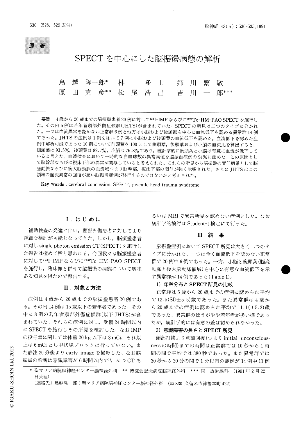
4歳から20歳までの脳振盪患者20例に対して123I-IMPならびに99mTc-HM-PAO SPECTを施行した。その内6例は若年者頭部外傷症候群(JHTS)が含まれていた。SPECTの所見は二つのタイプに分かれた。一つは血流異常を認めない正常群6例と他方は小脳および後頭部を中心に血流低下を認める異常群14例であった。JHTSの症例は1例を除いて7例に小脳および後頭葉の血流低下を認めた。血流低下を認めた症例中解析可能であった10例について前頭葉を100として側頭葉,後頭葉および小脳の血流比を算出すると,側頭葉は93.5%,後頭葉は82.7%,小脳は76書8%であり,統計学的に後頭葉と小脳は有意に血流が低下していると言えた。血液検査において一時的な白血球数の異常高値を脳振盪症例の94%に認めた。この原因として脳幹部ならびに視床下部の異常が関与していると考えられた。これらの所見から脳振蘯の責任病巣として脳底動脈ならびに後大脳動脈の血流域つまり脳幹部,視床下部の関与が強く示唆された。さらにJHTSはこの領域の血流異常の回復が悪い脳振盪症例が移行するのではないかと考えられた。
123I-IMP and Tc-PAO SPECT were performed in 20 cases of cerebral concussion ranging in age from 4 to 20 years old, including six cases of the juvenile haed trauma syndrome (JHTS). The SPECT findings were divided into two main types : six cases in the normal group with no blood flow abnor-malities, and 14 cases in abnormal group showing reduced blood flow, mainly in cerebellum and oc-cipital lobe except in one case. In 10 cases with reduced blood flow which could be analyzed, calcu-lation of the blood flow ratio in the temporal and occipital lobes and the cerebellum with the frontal lobe taken as 100 showed values of 93.5% for the temporal lobe, 82.7% for the occipital lobe and 76. 8% for the cerebellum. A statistically significant reduction in blood flow occurred in the occipital lobe and cerebellum. In blood examination, abnor-mally high values of white blood cell counts were observed transiently in 94% of cerebral concussion cases. Abnormalities in brain stem and hypoth-alamus appeared to cause these abnormal WBC values. From these findings, it was suggested that the blood flow regions of the basilar and posterior cerebral arteries, i. e., the brain stem and hypoth-alamus are closely connected with the lesions responsible for cerebral concussion. It also appear-ed that the JHTS occurs in cerebral concussion cases where recovery of the abnormal blood flow in these regions in poor.

Copyright © 1991, Igaku-Shoin Ltd. All rights reserved.


