Japanese
English
- 有料閲覧
PPV購入案内
1,320円 (1,200円+税10%) こちらは、99ページから105ページの文献です。購入には医書.jp本会員登録が必要です。
会員登録完了後に改めて上記「購入サイトへ」を選択してください。
または「購入サイトへ」選択後に「購入ログイン」、「新規会員登録」の順に進んでください。
会員登録について詳しくはをご確認ください。
- Abstract 文献概要
- 1ページ目 Look Inside
はじめに
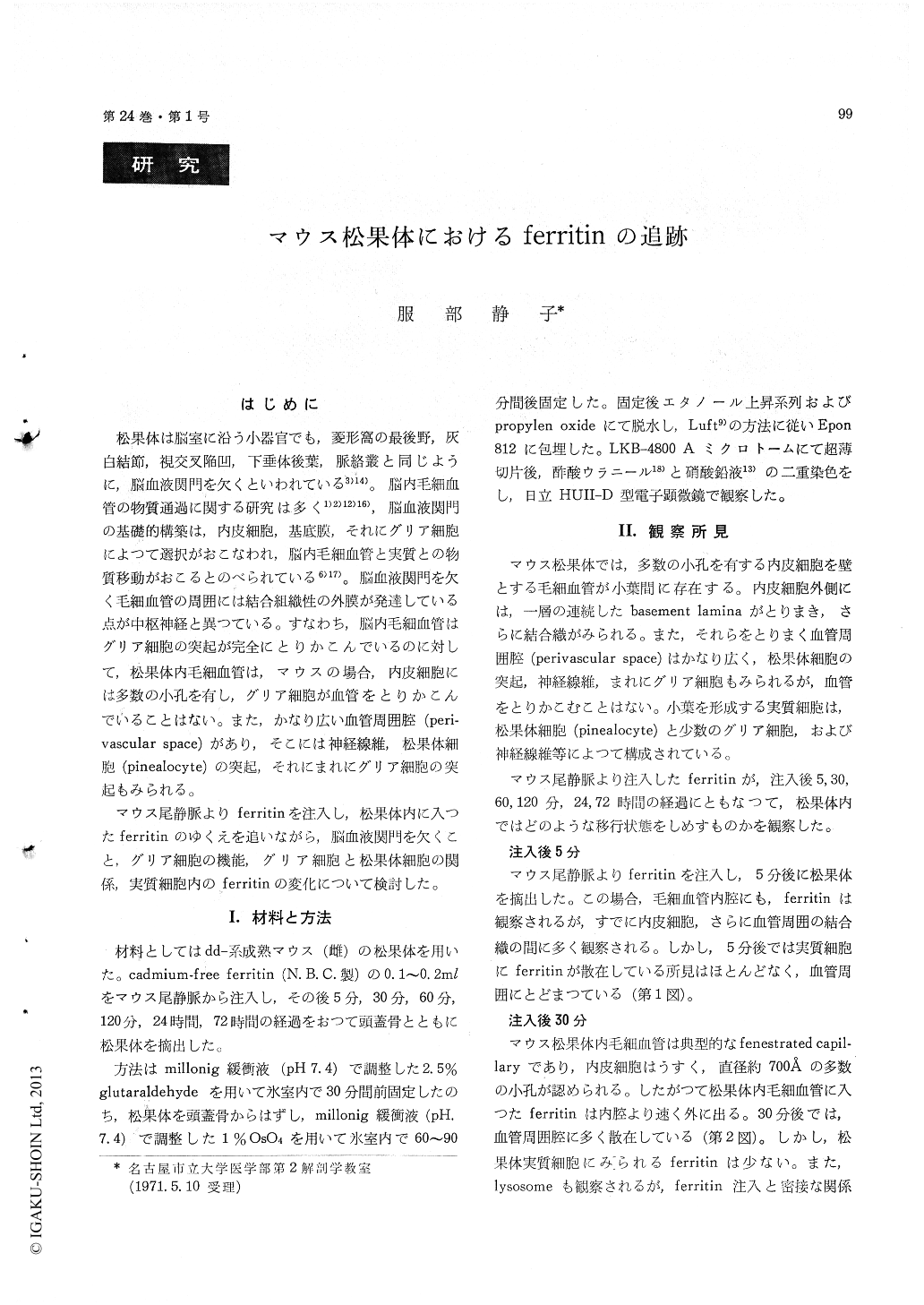
松果体は脳室に沿う小器官でも,菱形窩の最後野,灰白結節,視交叉陥凹,下垂体後葉,脈絡叢と同じように,脳血液関門を欠くといわれている3)14)。脳内毛細血管の物質通過に関する研究は多く1)2)12)16),脳血液関門の基礎的構築は,内皮細胞,基底膜,それにグリア細胞によつて選択がおこなわれ,脳内毛細血管と実質との物質移動がおこるとのべられている6)17)。脳血液関門を欠く毛細血管の周囲には結合組織性の外膜が発達している点が中枢神経と異つている。すなわち,脳内毛細血管はグリア細胞の突起が完全にとりかこんでいるのに対して,松果体内毛細血管は,マウスの場合,内皮細胞には多数の小孔を有し,グリア細胞が血管をとりかこんでいることはない。また,かなり広い血管周囲腔(peri—vascular space)があり,そこには神経線維,松果体細胞(pinealocyte)の突起,それにまれにグリア細胞の突起もみられる。
マウス尾静脈よりferritinを注入し,松果体内に入つたferritinのゆくえを追いながら,脳血液関門を欠くこと,グリア細胞の機能,グリア細胞と松果体細胞の関係,実質細胞内のferritinの変化について検討した。
1. In the capillaries of the mouse pineal bodythe endothelial cells possess numerous fenestrationsaveraging 700 Å in diameter. The capillaries aresurrounded by wide perivascular space instead ofcomplete glial investment as in the central nervoussystem. Pinealocyte processes, nerve fibers andoccasional glial cell processes occur in the space.
2. At 5 minuites after intravenous injection fer-ritin particles are seen outside of the vessels.
3. At 30 minuites after injection ferritin particlesare seen in the perivascular space as well as inthe parencymal cells (pinealocytes and glial cells).
4. At 60 minuites the particles are seen abundantlyin the parencymal cells which show an increase inprimary lysosomes at this stage.
5. At 120 minuites scattered ferritin particles inthe cytoplasm aggregate to form phagosomes orsecondary lysosomes. This change is more noti-ceable at 24 to 72 hours.
6. Glial cells in the mouse pineal body do notshow a function as a barrier as in the central nerv-ous system.
Copyright © 1972, Igaku-Shoin Ltd. All rights reserved.