Japanese
English
- 有料閲覧
PPV購入案内
1,320円 (1,200円+税10%) こちらは、179ページから182ページの文献です。購入には医書.jp本会員登録が必要です。
会員登録完了後に改めて上記「購入サイトへ」を選択してください。
または「購入サイトへ」選択後に「購入ログイン」、「新規会員登録」の順に進んでください。
会員登録について詳しくはをご確認ください。
- Abstract 文献概要
- 1ページ目 Look Inside
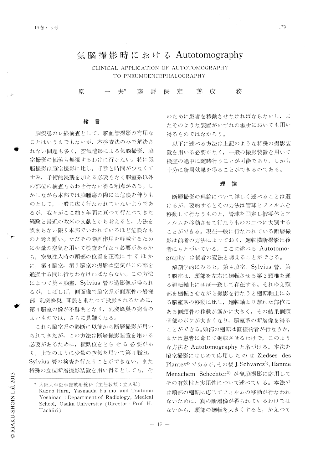
脳室,気脳撮影時のAutotornographyについてその理論,方法について述べ,第4脳室,Sylvius管,第3脳室,トルコ鞍部の診断に有効なことは証明し得たので,2〜3の具体例を例示した。
The fourth ventricle and the aqueduct of Sylvius can be shown well after injection of only a small amount of air, but these are sometimes obscured by the shadow of the pet-rous bone, especially marked by the well-developed mastoid cells.
Anatomically, the fourth ventricle and aque-duct of Sylvius are on the rotation axis of the patient's head, and, therefore, details of these can be obtained from the tomographic lateral view, by means of rotating the pati-ent's head from side to side. Rotating angle should be controlled within 20 to 30 degrees for the fourth ventricle and aqueduct of Syl-vius, and within the limits of 10 degrees for the third ventricle and suprasellar region. X-ray exposure must be made during the rota-tion of the patient's head. Exposure time varies between 1 and 3 seconds.
This method can be used with advantage to show structures that are obscured in sim-ple pneumoencephalograms, and offers most help in abnormalities in the region of aqueduct and fourth ventricle.
Autotomography facilitates diagnosis and also aids in assessing operative interventions.
Copyright © 1962, Igaku-Shoin Ltd. All rights reserved.