Japanese
English
- 有料閲覧
- Abstract 文献概要
- 1ページ目 Look Inside
I.はじめに
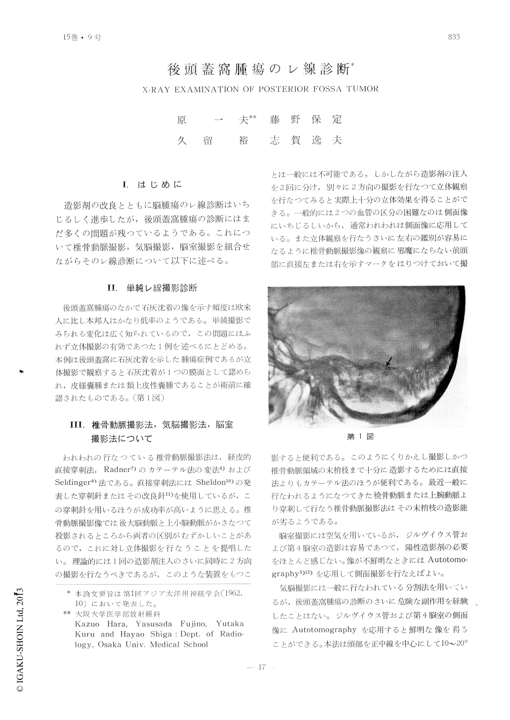
造影剤の改良とともに脳腫瘍のレ線診断はいちじるしく進歩したが,後頭蓋窩腫瘍の診断にはまだ多くの問題が残つているようである。これについて椎骨動脈撮影,気脳撮影,脳室撮影を組合せながらそのレ線診断について以下に述べる。
Cerebellar tumor.
In vertebral angiography signs listed as in-dicative of cerebellar tumour are;
(1) presence of pathologic blood-vessels in the cerebellar region;
(2) elevation and stretching of the superior cerebellar artery;
(3) absence of blood-vessels in the cerebe-llar portion;
(4) stretching of the posterior inferior cere-bellar artery; and
(5) displacement of the medical branch of the posterior inferior cerebellar artery to the opposite side.
As the shadows of the superior cerebellar arteries are usually superimposed on the pos-terior cerebral arteries, stereoscopic films are superior for differentiating them. In our ex-perience, pneumoventriclography is superior to vertebral angiography in that it gives an accurate location of the tumor, whereas ver-tebral angiography is superior to pneumoven-triculography in that nature of the tumor can be determind. Usually by pneumoencephalog-raphy, only non-specific tonsillar herniation can be proved.
Tumors of the brain stem and the fourth ventricle.
The most reliable signs of these tumors are observed by pneumoencephalography as well as pneumoventriculography. Pneumoen-cephalography is superior to peumoventricu-lography in that pontine cistern and medullary cistern can be visualized. However, it is sometimes impossible to demonstrate the ven-tricular system by this means. The fourth ventricle and Sylvian duct in lateral projection are sometimes obscured by the shadows of the petrous bones. It is advisable, therefore, to take an autotomogram by moving the pa-tient's head to both sides at an angle of 10 degrees. By this means the ventricle, even in a small amount of air, can be clearly demon-strated.
Extracerebral tumor.
In addition to cisternography, pneumoence-phalography is the roentgenologic procedure of choice in diagnosing extracerebral tumor. By concurrent use of these procedures, it is possible to arrive at an accurate appreciation of the actual size and contour of the tumor, even if it is a small tumor which does not occur any displacement and deformation ofthe ventricular system. This is also true in a case of trigeminal neurinoma which arises immediately beneath the anterolateral portion of the incisura tentoril. By vertebral angiog-ram it can only be shown that the tumor produces a slight upward displacement of the superior cerebellar artery on the affected side. Thus, pneumoencephalography is considered to be superior to vertebral angiography.
Cerebellopontine angle tumor can also be demonstrated by pneumoencephalography whi-ch is found to be useful in confirming the tumor arising from bilateral angles. When specific changes in vertebral angiography and plain roentgenogram are not present, there is little possibility of distinguishing this tumor from ones such as neurinoma, meningioma and dermoid cyst. In vertebral angiography the following signs are liable to occur : disp-lacementof the basilar artery to the opposite side ; stretching of the internal auditory artery and other small arteries ; elevation of the superior cerebellar artery as the tumor grows in size, posterior cerebral artery is elevated, peduncular fork is deformed and there is an avascular area in upper portion of the petrous bones. In neurinoma mesh-like vessels may be seen. In a small tumor pneumoencephalog-raphy displays further details of the tumor.
Clival tumor.
In vertebral angiography the basilar artery is displaced posteriorly or to the opposite side by the tumor mass, but by pneumoencepha-lography the contour and extension of the tumor can be demonstrated.

Copyright © 1963, Igaku-Shoin Ltd. All rights reserved.


