Japanese
English
- 有料閲覧
PPV購入案内
1,320円 (1,200円+税10%) こちらは、1043ページから1054ページの文献です。購入には医書.jp本会員登録が必要です。
会員登録完了後に改めて上記「購入サイトへ」を選択してください。
または「購入サイトへ」選択後に「購入ログイン」、「新規会員登録」の順に進んでください。
会員登録について詳しくはをご確認ください。
- Abstract 文献概要
- 1ページ目 Look Inside
- 参考文献 Reference
- サイト内被引用 Cited by
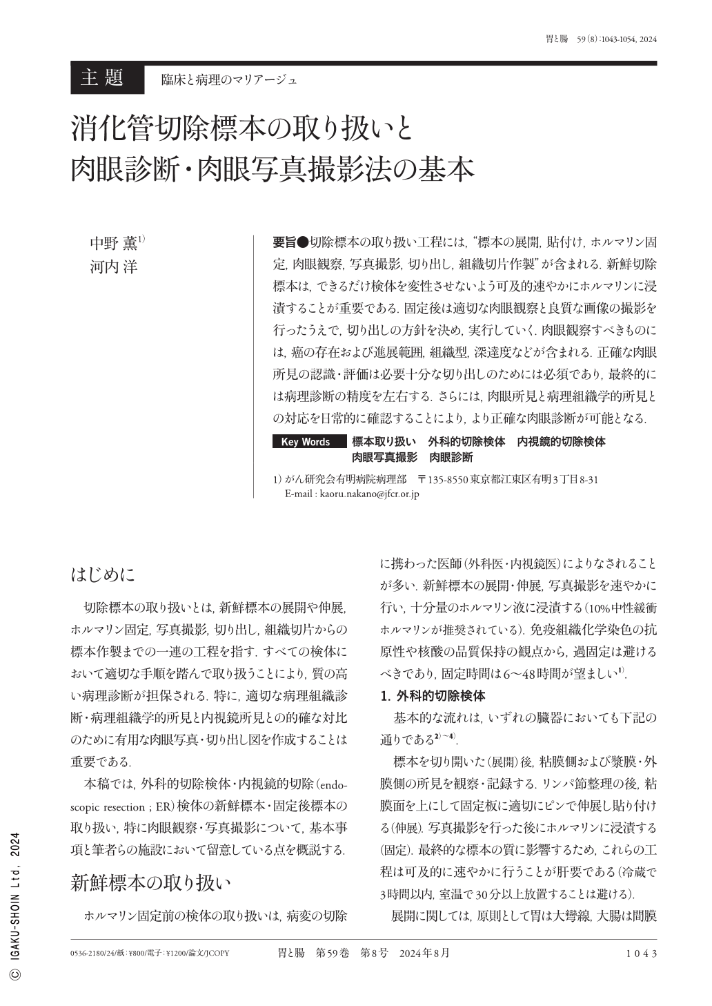
要旨●切除標本の取り扱い工程には,“標本の展開,貼付け,ホルマリン固定,肉眼観察,写真撮影,切り出し,組織切片作製”が含まれる.新鮮切除標本は,できるだけ検体を変性させないよう可及的速やかにホルマリンに浸漬することが重要である.固定後は適切な肉眼観察と良質な画像の撮影を行ったうえで,切り出しの方針を決め,実行していく.肉眼観察すべきものには,癌の存在および進展範囲,組織型,深達度などが含まれる.正確な肉眼所見の認識・評価は必要十分な切り出しのためには必須であり,最終的には病理診断の精度を左右する.さらには,肉眼所見と病理組織学的所見との対応を日常的に確認することにより,より正確な肉眼診断が可能となる.
Handling resected specimens includes gastrointestinal specimen opening and stretching, formalin fixation, macroscopic observation, photography, sampling, and tissue section preparation. Accurately assessing the macroscopic findings and taking high-quality photographs to identify and execute tissue sampling for fixed specimens is crucial. Macroscopic diagnosis includes evaluating the presence and extent of cancer, histological type, and invasion depth. Accurate macroscopic finding evaluation is essential for sufficient tissue sampling and significantly affects the final pathological diagnosis. Understanding the macroscopic findings that need to be observed and regularly confirming their correspondence with histological findings enable more accurate macroscopic diagnosis. Additionally, an accurate understanding of macroscopic findings is important when comparing them with endoscopic and radiological findings.
Copyright © 2024, Igaku-Shoin Ltd. All rights reserved.