Japanese
English
- 有料閲覧
- Abstract 文献概要
- 1ページ目 Look Inside
- 参考文献 Reference
- サイト内被引用 Cited by
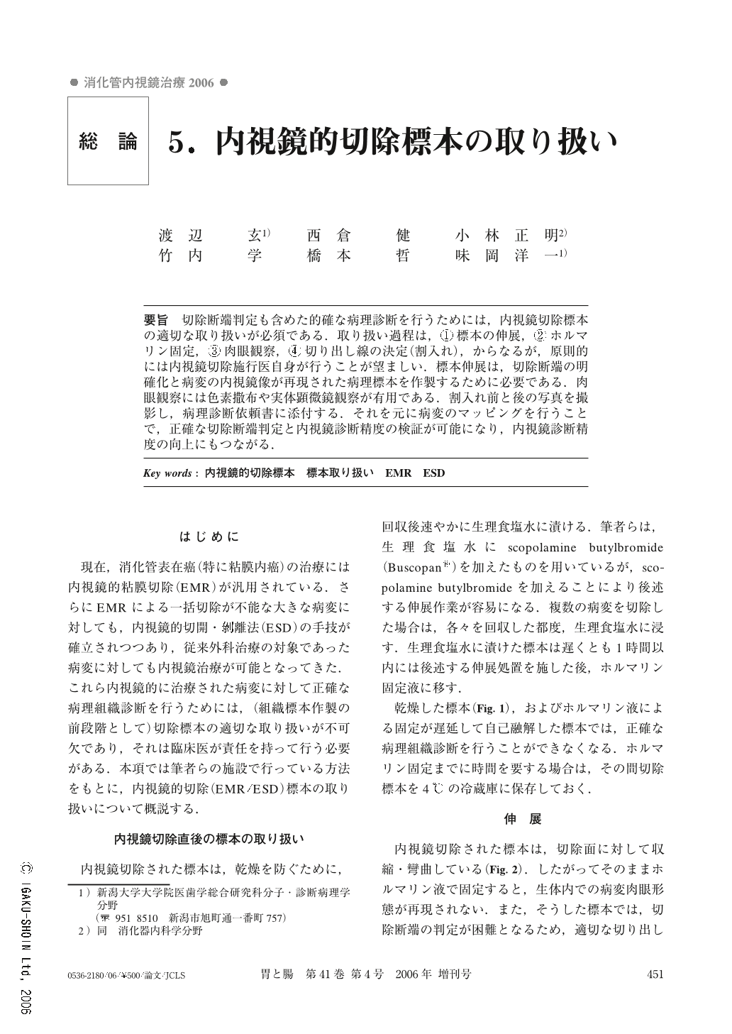
要旨 切除断端判定も含めた的確な病理診断を行うためには,内視鏡切除標本の適切な取り扱いが必須である.取り扱い過程は,①標本の伸展,②ホルマリン固定,③肉眼観察,④切り出し線の決定(割入れ),からなるが,原則的には内視鏡切除施行医自身が行うことが望ましい.標本伸展は,切除断端の明確化と病変の内視鏡像が再現された病理標本を作製するために必要である.肉眼観察には色素撒布や実体顕微鏡観察が有用である.割入れ前と後の写真を撮影し,病理診断依頼書に添付する.それを元に病変のマッピングを行うことで,正確な切除断端判定と内視鏡診断精度の検証が可能になり,内視鏡診断精度の向上にもつながる.
Proper gross handling of endoscopically resected〔EMR (endoscopic mucosal resection) and ESD (endoscopic submucosal dissection)〕specimens is necessary for appropriate histopathological diagnosis for the purpose of assessing the nature and resected margin of a tumor, and for providing information necessary for further management of patients undergoing resection. Gross handling starts with adequate extension of the specimen, followed by formalin fixation and macroscopic and dye-aided stereomicroscopic observation. After detailed macroscopic observation, one should provide marking of the sectioning lines by a razor to indicate to the pathologist the need to make proper histological sections. Macroscopic photographs of the specimen should be taken both before and after the marking of the sectioning lines. It is recommended to send prints of these to the pathologist with the clinical report, so that histological mapping of the tumor can be made. The mapping is necessary for precise judgment of the resected margin, and to verify the appropriateness of the endoscopic diagnosis. In principle, all these procedures are recommended to be done by the endoscopist who has carried out the EMR or ESD. However, it is permitted to ask the pathologist to perform the procedures after the fixation, when there is good mutual communication between the endoscopist and the pathologist. It is emphasized that proper gross handling of the specimen is not only necessary for appropriate histopathological assessing, but also for the improvement of the precision of the endoscopic diagnosis.

Copyright © 2006, Igaku-Shoin Ltd. All rights reserved.


