Japanese
English
- 有料閲覧
- Abstract 文献概要
- 1ページ目 Look Inside
- サイト内被引用 Cited by
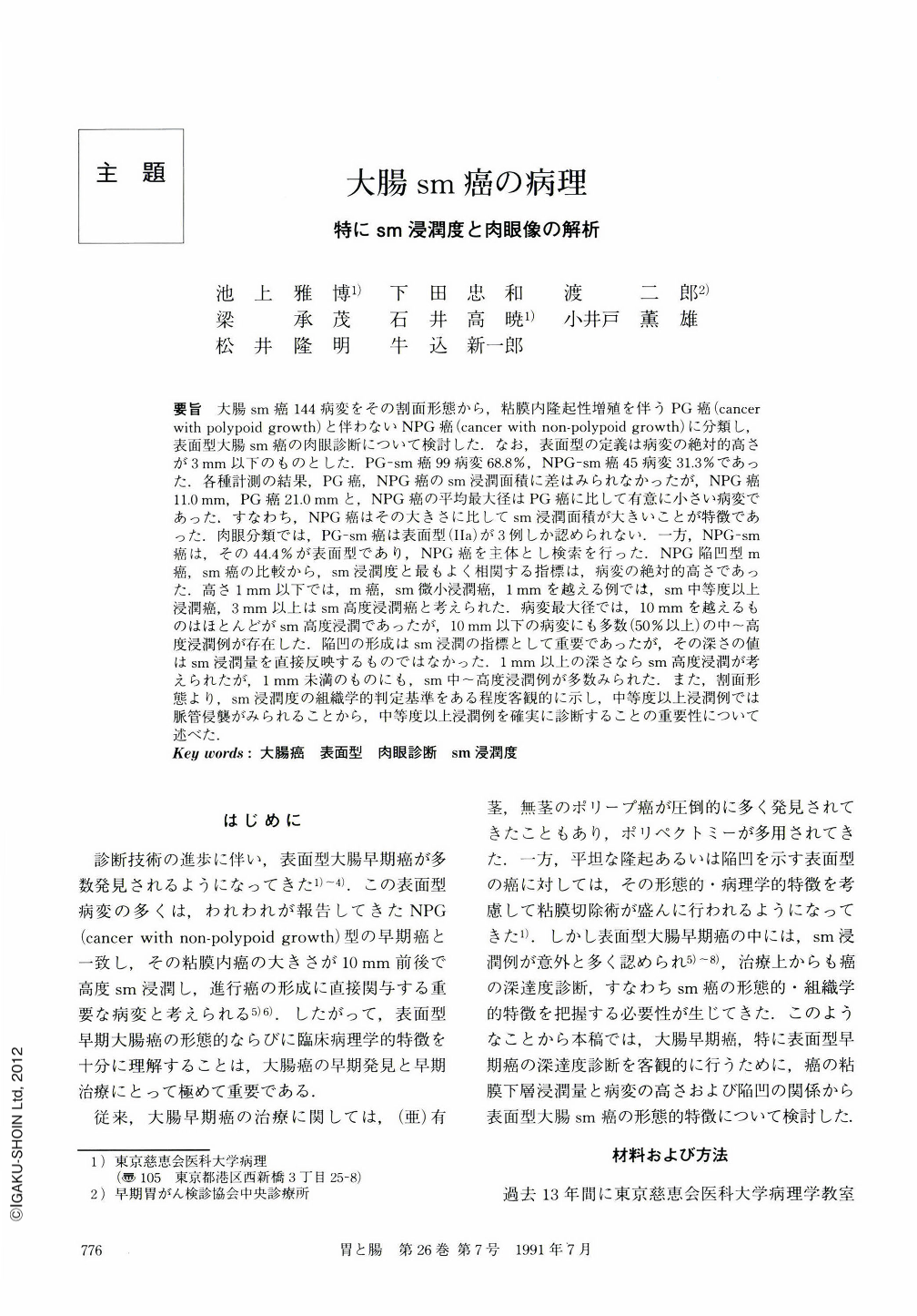
要旨 大腸sm癌144病変をその割面形態から,粘膜内隆起性増殖を伴うPG癌(cancer wlth polypoid growth)と伴わないNPG癌(cancer with non-polypoid growth)に分類し,表面型大腸sm癌の肉眼診断について検討した.なお,表面型の定義は病変の絶対的高さが3mm以下のものとした.PG-sm癌99病変68.8%,NPG-sm癌45病変31.3%であった.各種計測の結果,PG癌,NPG癌のsm浸潤面積に差はみられなかったが,NPG癌11.Omm,PG癌21.Ommと,NPG癌の平均最大径はPG癌に比して有意に小さい病変であった.すなわち,NPG癌はその大きさに比してsm浸潤面積が大きいことが特徴であった.肉眼分類では,PG-sm癌は表面型(IIa)が3例しか認められない.一方,NPG-sm癌は,その44.4%が表面型であり,NPG癌を主体とし検索を行った.NPG陥凹型m癌,sm癌の比較から,sm浸潤度と最もよく相関する指標は,病変の絶対的高さであった.高さ1mm以下では,m癌,sm微小浸潤癌,1mmを越える例では,sm中等度以上浸潤癌,3mm以上はsm高度浸潤癌と考えられた.病変最大径では,10mmを越えるものはほとんどがsm高度浸潤であったが,10mm以下の病変にも多数(50%以上)の中~高度浸潤例が存在した.陥凹の形成はsm浸潤の指標として重要であったが,その深さの値はsm浸潤量を直接反映するものではなかった.1mm以上の深さならsm高度浸潤が考えられたが,1mm未満のものにも,sm中~高度浸潤例が多数みられた.また,割面形態より,sm浸潤度の組織学的判定基準をある程度客観的に示し,中等度以上浸潤例では脈管侵襲がみられることから,中等度以上浸潤例を確実に診断することの重要性について述べた.
Macroscopical diagnosis of superficial colorectal cancer which showed submucosal invasion was studied. Superficial colorectal cancer was defined as that the absolute height of the lesion was less than 3 mm. Colorectal cancers were classified into two groups:Those accompanied by intramucosal polypoid growth (PG-ca) and those with non-polypoid growth (NPG-ca). Ninety nine lesions (68.8%) were PG-ca, while 45 lesions (31.3%) were NPG-ca. In the measurement value of the area of invasive cancer tissue in the submucosal layer, there was no difference between PG-ca and NPG-ca.
On the other hand, however, the average maximum diameter of PG-ca was 21.O mm, while the lesions of NPG-ca with an average diameter of 11.O mm were smaller. In other words, compared with PG-ca, a characteristic of NPG-ca was that the invasive cancer area in the submucosal layer was quite large when compared with the maximum diameter of its lesion. In macroscopical classification,PG-ca showed only 3 lesions of the superficial type of cancer, while 44.4% of submucosal invasive NPG-ca were of the superficial type.
We then discussed the fact that to make a diagnosis about the depth of invasion of NPG-ca, a morphometrical study of intramucosal and submucosal invasive NPG-ca can be of use. There was a definite correlation between the absolute height of the lesion and the degree of submucosal invasion. The heights of cancerous lesions were categorized as follows:Less than 1 mm in height-intramucosal cancer or minimal submucosal invasive cancer. Over 1 mm in height-moderate or massive submucosal invasive cancer. More than 3 mm in height-massive submucosal invasive cancer. The maximum diameter of lesions over 10 mm, usually indicated massive submucosal invasion. Many of the lesions less than 10 mm in maximum diameterindicated moderate or massive submucosal invasion. Deep depression was important evidence of invasion into the submucosal layer. Depression over 1 mm in depth indicated massive submucosal invasion. As one result of this study, we found that cancer with minimal invasion of the submucosal layer did not result in lymphatic and venous permeation. To determine whether there is moderate or massive invasion into the submucosal layer, it is important to make exact macroscopical diagnosis.

Copyright © 1991, Igaku-Shoin Ltd. All rights reserved.


