Japanese
English
- 有料閲覧
- Abstract 文献概要
- 1ページ目 Look Inside
- 参考文献 Reference
- サイト内被引用 Cited by
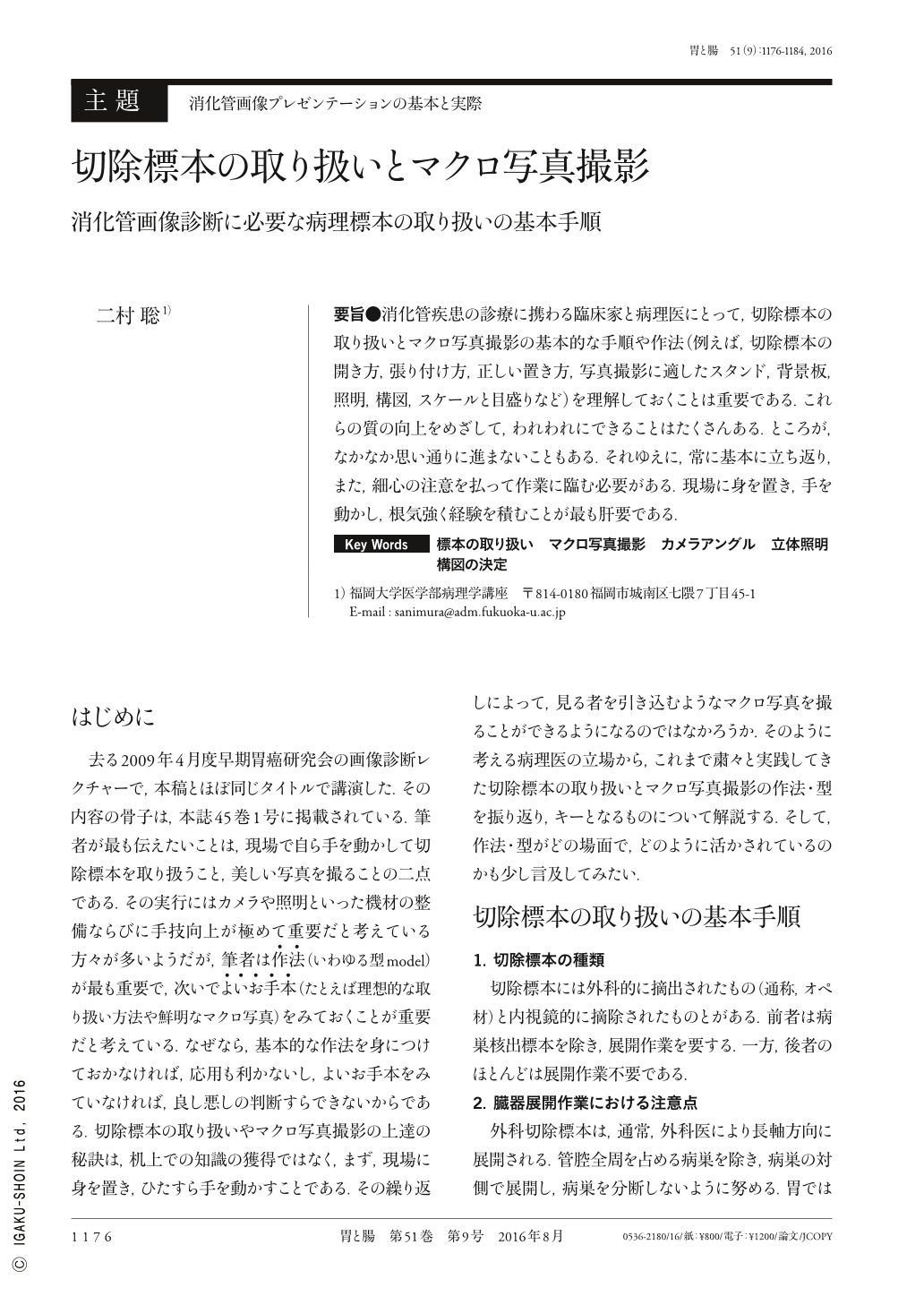
要旨●消化管疾患の診療に携わる臨床家と病理医にとって,切除標本の取り扱いとマクロ写真撮影の基本的な手順や作法(例えば,切除標本の開き方,張り付け方,正しい置き方,写真撮影に適したスタンド,背景板,照明,構図,スケールと目盛りなど)を理解しておくことは重要である.これらの質の向上をめざして,われわれにできることはたくさんある.ところが,なかなか思い通りに進まないこともある.それゆえに,常に基本に立ち返り,また,細心の注意を払って作業に臨む必要がある.現場に身を置き,手を動かし,根気強く経験を積むことが最も肝要である.
It is important for clinicians and pathologists to understand the basic requirements for the successful photography of resected specimens. These include the anatomically correct orientation of the specimens, composition of the image, choice of optimal lighting conditions, and inclusion of appropriate scale markers. Forward planning of the facilities and equipment required using criteria discussed in this text should improve both the preparation and subsequent photography of the tissue specimens. Errors may occur despite such careful preparation and it is, therefore, important to pay close attention to the basic requirements at all times. I firmly believe that the accumulation of experience in the field is the easiest and best way to improve the preparation and photography of resected specimens.

Copyright © 2016, Igaku-Shoin Ltd. All rights reserved.


