Japanese
English
- 有料閲覧
PPV購入案内
1,320円 (1,200円+税10%) こちらは、246ページから253ページの文献です。購入には医書.jp本会員登録が必要です。
会員登録完了後に改めて上記「購入サイトへ」を選択してください。
または「購入サイトへ」選択後に「購入ログイン」、「新規会員登録」の順に進んでください。
会員登録について詳しくはをご確認ください。
- Abstract 文献概要
- 1ページ目 Look Inside
- 参考文献 Reference
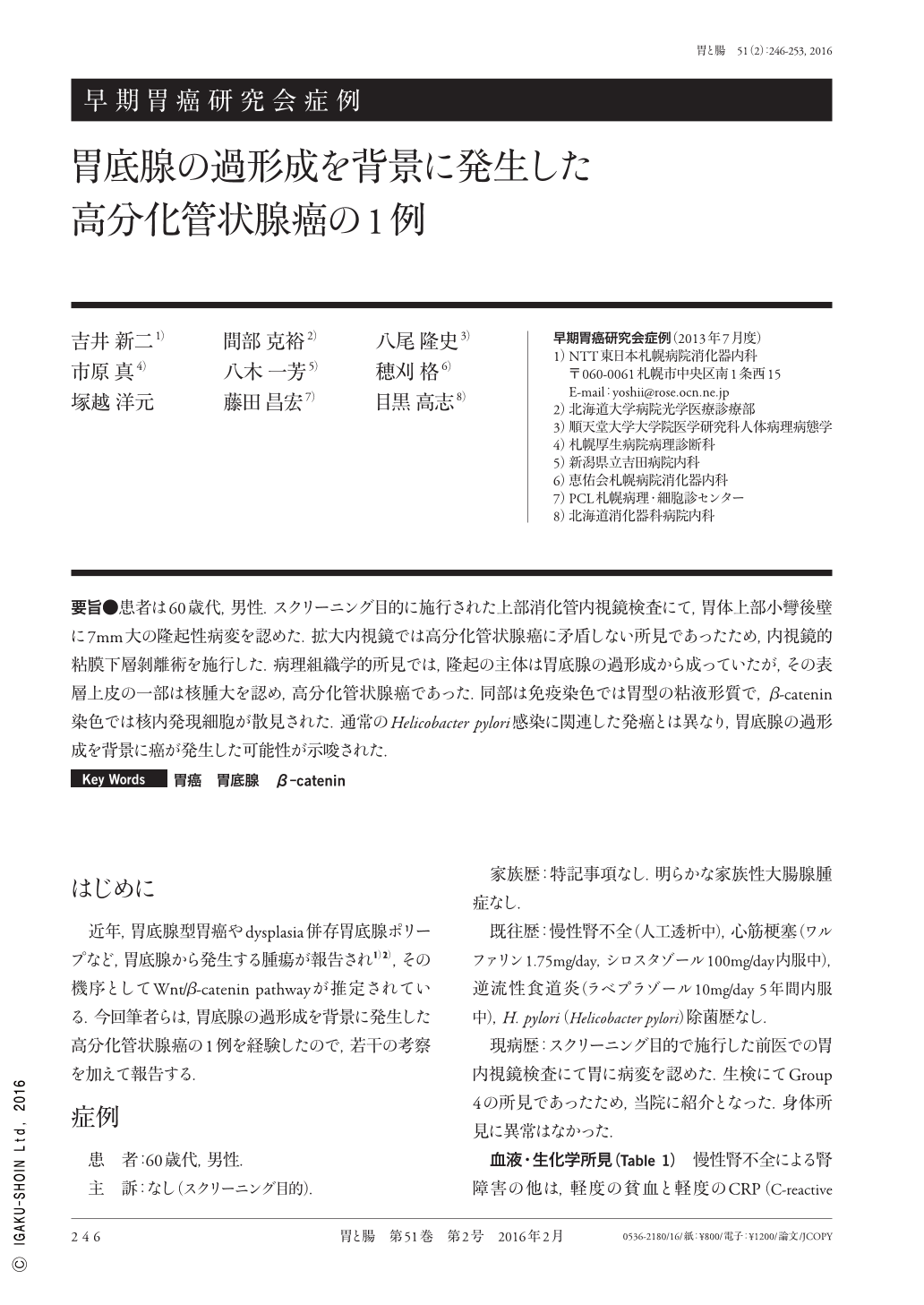
要旨●患者は60歳代,男性.スクリーニング目的に施行された上部消化管内視鏡検査にて,胃体上部小彎後壁に7mm大の隆起性病変を認めた.拡大内視鏡では高分化管状腺癌に矛盾しない所見であったため,内視鏡的粘膜下層剝離術を施行した.病理組織学的所見では,隆起の主体は胃底腺の過形成から成っていたが,その表層上皮の一部は核腫大を認め,高分化管状腺癌であった.同部は免疫染色では胃型の粘液形質で,β-catenin染色では核内発現細胞が散見された.通常のHelicobacter pylori感染に関連した発癌とは異なり,胃底腺の過形成を背景に癌が発生した可能性が示唆された.
The patient was a 60-year-old man who was found to have an elevated lesion 7mm in diameter in the posterior wall of the lesser curvature of the stomach by upper gastrointestinal endoscopy conducted for screening. Because the findings of magnifying endoscopy were consistent with a well-differentiated adenocarcinoma, endoscopic submucosal dissection was performed. Histopathologically, although most of the lesion showed fundic gland hyperplasia, a part of the superficial epithelium showed nucleolar hypertrophy, confirming a well-differentiated adenocarcinoma. Immunostaining of the lesion showed a gastric phenotype with some cells positive for nuclear β-catenin. Different from Helicobacter pylori-related cancer, the fundic gland hyperplasia was thought to be the underlying cause of cancer in this case.
Copyright © 2016, Igaku-Shoin Ltd. All rights reserved.