Japanese
English
- 有料閲覧
- Abstract 文献概要
- 1ページ目 Look Inside
- 参考文献 Reference
- サイト内被引用 Cited by
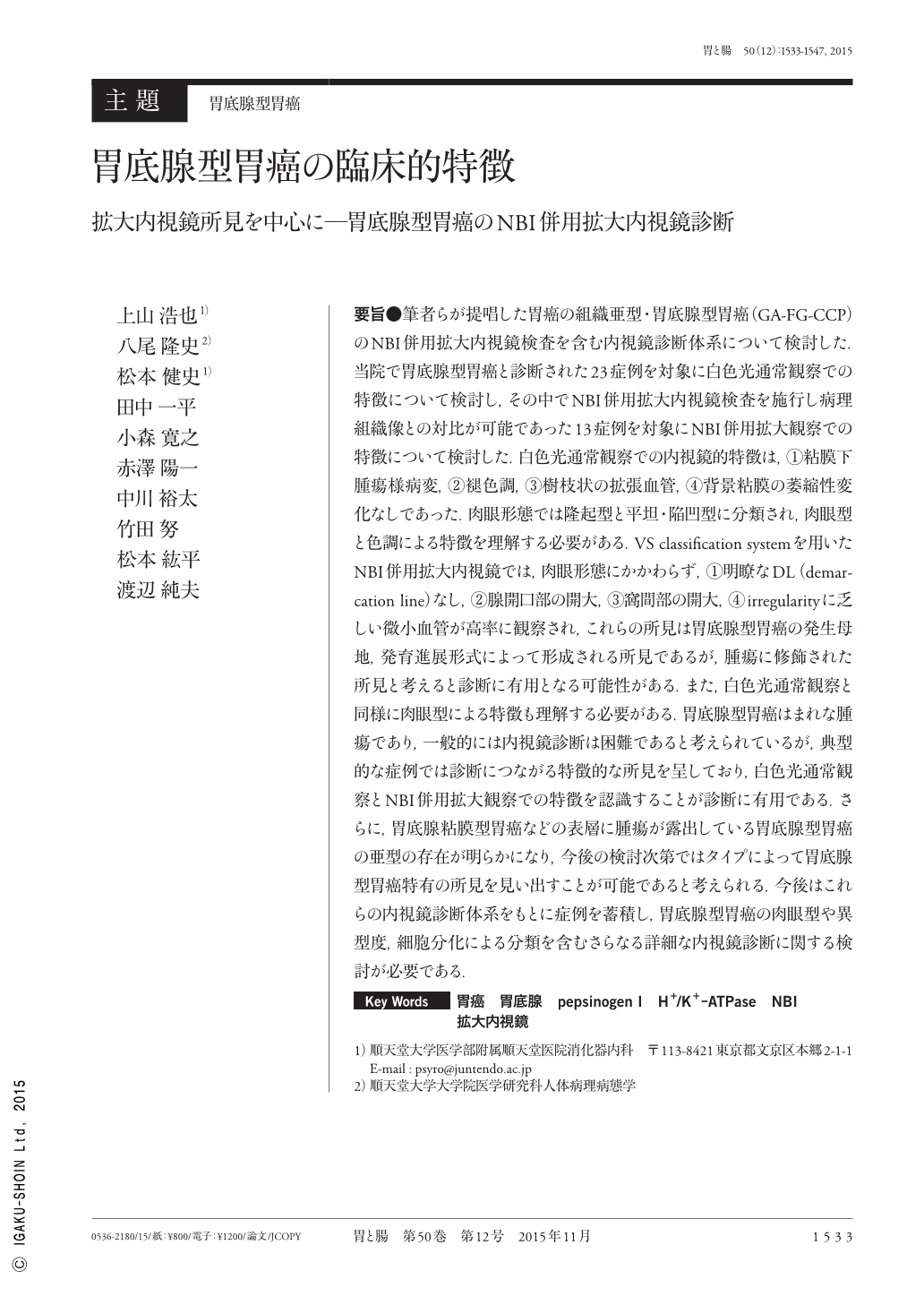
要旨●筆者らが提唱した胃癌の組織亜型・胃底腺型胃癌(GA-FG-CCP)のNBI併用拡大内視鏡検査を含む内視鏡診断体系について検討した.当院で胃底腺型胃癌と診断された23症例を対象に白色光通常観察での特徴について検討し,その中でNBI併用拡大内視鏡検査を施行し病理組織像との対比が可能であった13症例を対象にNBI併用拡大観察での特徴について検討した.白色光通常観察での内視鏡的特徴は,①粘膜下腫瘍様病変,②褪色調,③樹枝状の拡張血管,④背景粘膜の萎縮性変化なしであった.肉眼形態では隆起型と平坦・陥凹型に分類され,肉眼型と色調による特徴を理解する必要がある.VS classification systemを用いたNBI併用拡大内視鏡では,肉眼形態にかかわらず,①明瞭なDL(demarcation line)なし,②腺開口部の開大,③窩間部の開大,④irregularityに乏しい微小血管が高率に観察され,これらの所見は胃底腺型胃癌の発生母地,発育進展形式によって形成される所見であるが,腫瘍に修飾された所見と考えると診断に有用となる可能性がある.また,白色光通常観察と同様に肉眼型による特徴も理解する必要がある.胃底腺型胃癌はまれな腫瘍であり,一般的には内視鏡診断は困難であると考えられているが,典型的な症例では診断につながる特徴的な所見を呈しており,白色光通常観察とNBI併用拡大観察での特徴を認識することが診断に有用である.さらに,胃底腺粘膜型胃癌などの表層に腫瘍が露出している胃底腺型胃癌の亜型の存在が明らかになり,今後の検討次第ではタイプによって胃底腺型胃癌特有の所見を見い出すことが可能であると考えられる.今後はこれらの内視鏡診断体系をもとに症例を蓄積し,胃底腺型胃癌の肉眼型や異型度,細胞分化による分類を含むさらなる詳細な内視鏡診断に関する検討が必要である.
Gastric adenocarcinoma of fundic gland type(chief cell predominant type ; GA-FG-CCP)has recently been proposed as a new, rare variant of gastric adenocarcinoma. The aim of the current study was to evaluate the endoscopic diagnostic system of GA-FG-CCP using ME-NBI(magnifying endoscopy with narrow-band imaging). The endoscopic features of GA-FG-CCP were analyzed using CE(conventional endoscopy)and ME-NBI in 23 cases. The most common features observed with CE were as follows : 1)submucosal tumor shape, 2)whitish color, 3)dilated vessels with branching architecture, and 4)surrounding mucosa without atrophic changes. Endoscopic findings of GA-FG-CCP with ME-NBI could not meet the criteria for diagnosis of carcinoma. However, the four most frequent features detected using ME-NBI were as follows : 1)indistinct line of demarcation between the lesion and the surrounding mucosa, 2)dilatation of the crypt opening, 3)dilatation of the intervening portion between the crypts, and 4)microvessels without distinct irregularities. These findings appear due to the location of tumor origin and the congestion caused by pressure from the tumor. The endoscopic diagnosis of GA-FG-CCP can be established by recognizing these endoscopic features using both CE and ME-NBI. To elucidate the natural history of GA-FG-CCP by assessing the classification based on the macroscopic findings, grade of atypia, and cell differentiation, further investigations should include cases with these endoscopic features and an accurate pathological diagnosis of GA-FG-CCP.

Copyright © 2015, Igaku-Shoin Ltd. All rights reserved.


