Japanese
English
- 有料閲覧
PPV購入案内
1,320円 (1,200円+税10%) こちらは、175ページから187ページの文献です。購入には医書.jp本会員登録が必要です。
会員登録完了後に改めて上記「購入サイトへ」を選択してください。
または「購入サイトへ」選択後に「購入ログイン」、「新規会員登録」の順に進んでください。
会員登録について詳しくはをご確認ください。
- Abstract 文献概要
- 1ページ目 Look Inside
- 参考文献 Reference
- サイト内被引用 Cited by
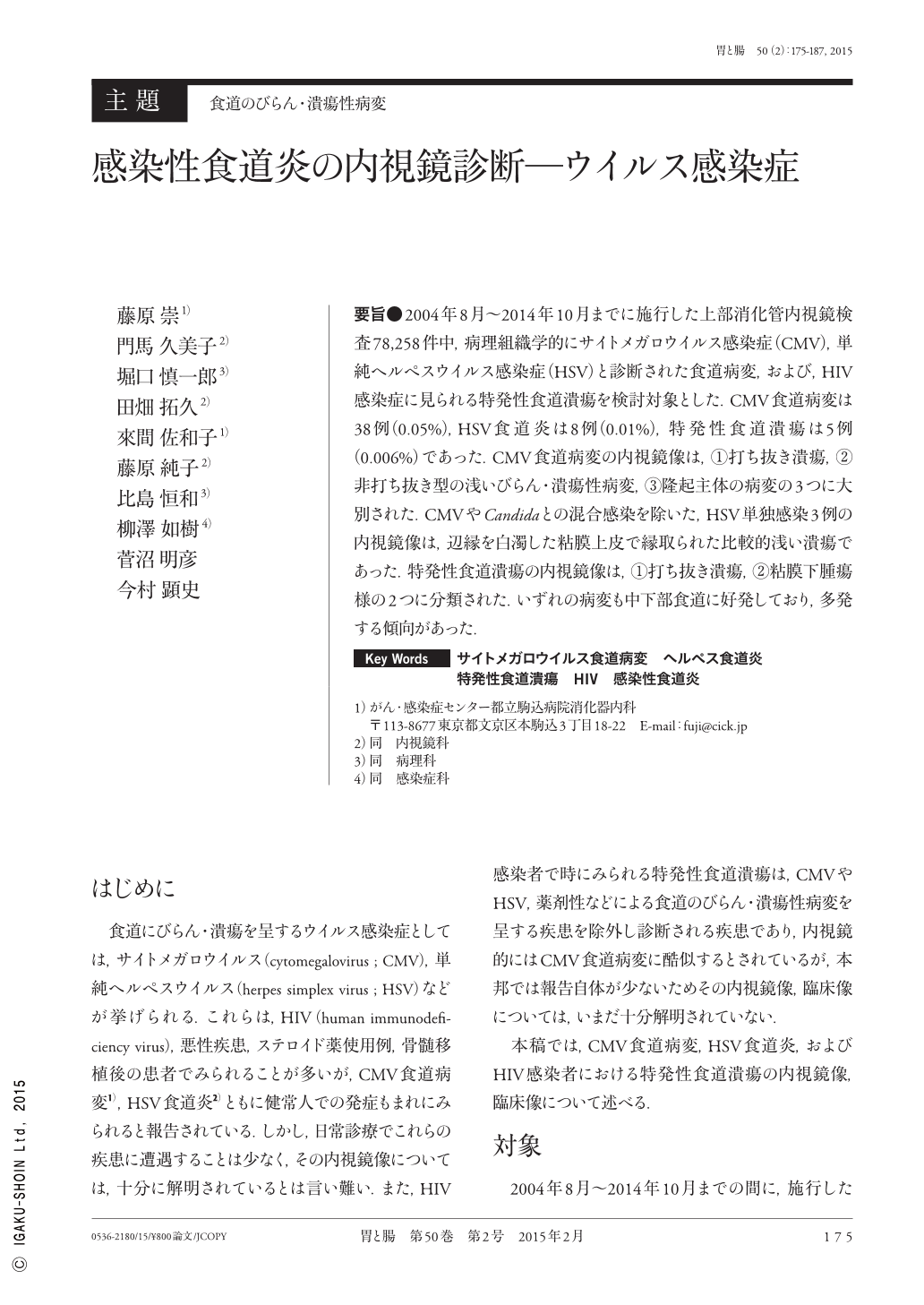
要旨●2004年8月〜2014年10月までに施行した上部消化管内視鏡検査78,258件中,病理組織学的にサイトメガロウイルス感染症(CMV),単純ヘルペスウイルス感染症(HSV)と診断された食道病変,および,HIV感染症に見られる特発性食道潰瘍を検討対象とした.CMV食道病変は38例(0.05%),HSV食道炎は8例(0.01%),特発性食道潰瘍は5例(0.006%)であった.CMV食道病変の内視鏡像は,(1)打ち抜き潰瘍,(2)非打ち抜き型の浅いびらん・潰瘍性病変,(3)隆起主体の病変の3つに大別された.CMVやCandidaとの混合感染を除いた,HSV単独感染3例の内視鏡像は,辺縁を白濁した粘膜上皮で縁取られた比較的浅い潰瘍であった.特発性食道潰瘍の内視鏡像は,(1)打ち抜き潰瘍,(2)粘膜下腫瘍様の2つに分類された.いずれの病変も中下部食道に好発しており,多発する傾向があった.
We investigated lesions of the esophagus diagnosed histopathologically as cytomegalovirus(CMV)and herpes simplex virus(HSV), and idiopathic esophageal ulcers thought to be related to human immunodeficiency virus(HIV)infection identified from a total of 78,258 upper gastrointestinal endoscopy procedures performed between August 2004 and October 2014. There were 38 cases(0.05%)of CMV esophageal lesion, eight cases(0.01%)of HSV esophagitis, and five cases(0.006%)of idiopathic esophageal ulcer. Endoscopic images of CMV esophageal lesions were broadly divided into the following three groups:erosive ulcer, non-erosive superficial ulceration/ulcerative lesion, and protruding lesion. Endoscopic images of three cases of HSV, excluding those with mixed infection of CMV and candida, indicated relatively superficial ulcers fringed with mucosal epithelium with whitish margins. Endoscopic images of idiopathic esophageal ulcers were categorized as either erosive ulcers or submucosal tumor-like lesions. All lesions were frequently found in the center and lower esophagus and tended to exhibit multiple onset.
Copyright © 2015, Igaku-Shoin Ltd. All rights reserved.