Japanese
English
- 有料閲覧
- Abstract 文献概要
- 1ページ目 Look Inside
- 参考文献 Reference
- サイト内被引用 Cited by
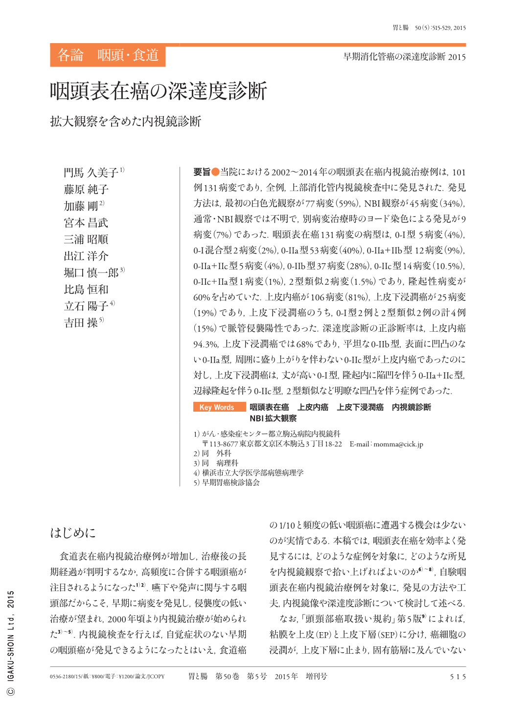
要旨●当院における2002〜2014年の咽頭表在癌内視鏡治療例は,101例131病変であり,全例,上部消化管内視鏡検査中に発見された.発見方法は,最初の白色光観察が77病変(59%),NBI観察が45病変(34%),通常・NBI観察では不明で,別病変治療時のヨード染色による発見が9病変(7%)であった.咽頭表在癌131病変の病型は,0-I型 5病変(4%),0-I混合型2病変(2%),0-IIa型53病変(40%),0-IIa+IIb型 12病変(9%),0-IIa+IIc型5病変(4%),0-IIb型37病変(28%),0-IIc型14病変(10.5%),0-IIc+IIa型1病変(1%),2型類似2病変(1.5%)であり,隆起性病変が60%を占めていた.上皮内癌が106病変(81%),上皮下浸潤癌が25病変(19%)であり,上皮下浸潤癌のうち,0-I型2例と2型類似2例の計4例(15%)で脈管侵襲陽性であった.深達度診断の正診断率は,上皮内癌94.3%,上皮下浸潤癌では68%であり,平坦な0-IIb型,表面に凹凸のない0-IIa型,周囲に盛り上がりを伴わない0-IIc型が上皮内癌であったのに対し,上皮下浸潤癌は,丈が高い0-I型,隆起内に陥凹を伴う0-IIa+IIc型,辺縁隆起を伴う0-IIc型,2型類似など明瞭な凹凸を伴う症例であった.
We examined 101 superficial pharyngeal cancer patients with 131 lesions treated by endoscopy between 2002 and 2014. In all patients, the lesions were detected during endoscopy of the upper gastrointestinal tract. The detection method included initial white-light imaging in 77 lesions(59%)and NBI(narrow-band imaging)in 45 lesions(34%). In the remaining nine lesions(7%)that were unclear on conventional NBI, the lesions were detected by iodine staining upon treatment of another lesion. The classification of 131 superficial pharyngeal cancer lesions included type 0-I in five lesions(4%), mixed type 0-I in two lesions(2%), type 0-IIa in 53 lesions(40%), type 0-IIa+IIb in 12 lesions(9%), type 0-IIa+IIc in five lesions(4%), type 0-IIb in 37 lesions(28%), type 0-IIc in 14 lesions(10.5%), type 0-IIc+IIa in one lesion(1%), and type 2-like in two lesions(1.5%), with protruding lesions accounting for 60%. According to the invasion depth, we found intraepithelial carcinomas in 106 lesions(81%)and subepithelial invasion in 25 lesions(19%). Intraepithelial carcinomas included flat type IIb, type IIa without surface irregularities, and type IIc without surrounding elevation, whereas cancer with subepithelial invasion included type 0-I with high elevation, type 0-IIa+IIc with depressed area in an elevated lesion, type 0-IIc with marginal elevation, and lesions with clear irregularities, such as type 2-like. In cancers with subepithelial invasion, positive vascular invasion was observed in a total of four patients(15%), including two patients with type 0-I lesions and two patients with type 2-like lesions.

Copyright © 2015, Igaku-Shoin Ltd. All rights reserved.


