Japanese
English
- 有料閲覧
- Abstract 文献概要
- 1ページ目 Look Inside
- 参考文献 Reference
要 旨
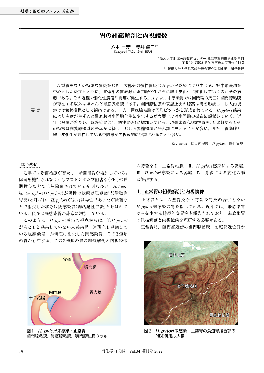
A型胃炎などの特殊な胃炎を除き,大部分の慢性胃炎はH.pylori感染により生じる。好中球浸潤を中心とした炎症とともに,胃体部の胃底腺が幽門腺化生さらに腸上皮化生に変化していくのがその病態である。その過程で消化性潰瘍や胃癌が発生する。H.pylori未感染胃では幽門輪の周囲に幽門腺粘膜が存在する以外はほとんど胃底腺粘膜である。幽門腺粘膜の表層上皮の腺窩は溝を形成し,拡大内視鏡では管状模様として観察できる。一方,胃底腺粘膜は円形ピットから形成されている。H.pylori感染により炎症が生ずると胃底腺は幽門腺化生に変化するが表層上皮は幽門腺の構造に類似していく。近年は除菌が普及し,既感染胃(非活動性胃炎)が増加している。現感染胃(活動性胃炎)と比較するとその特徴は非萎縮領域の発赤が消褪し,むしろ萎縮領域が発赤調に見えることが多い。また,胃底腺と腸上皮化生が混在している中間帯が内視鏡的に視認されることも多い。
In the majority of cases, chronic gastritis is caused by H.pylori infection; the exceptions are in cases of specialized gastritis, autoimmune gastritis, and so on. The pathology of chronic gastritis is as follows: fundic glands change to pyloric gland metaplasia and intestinal metaplasia with inflammation including that of neutrophils. The normal stomach without H.pylori infection consists of a fundic gland mucosa other than pyloric gland mucosa around the pyloric ring. The surface epithelium of the pyloric gland mucosa forms a gutter with crypts, resulting in a tubular pattern. On the other hand, the fundic gland mucosa forms round pits with crypts. As the inflammation of H.pylori transforms the fundic gland to a pyloric gland mucosa, the pattern of the surface epithelium changes from round pits to a tubular pattern, which is similar to that of the pyloric gland mucosa. In active gastritis, the fundic gland mucosa shows red color and an atrophic mucosa shows whitish color; however, in inactive gastritis, the fundic gland mucosa shows whitish color and the atrophic mucosa shows red color. Furthermore, an intermediate zone, which consist of a combination of fundic gland and intestinal metaplasia, becomes visible in inactive gastritis.

© tokyo-igakusha.co.jp. All right reserved.


