Japanese
English
- 有料閲覧
- Abstract 文献概要
- 1ページ目 Look Inside
- 参考文献 Reference
- サイト内被引用 Cited by
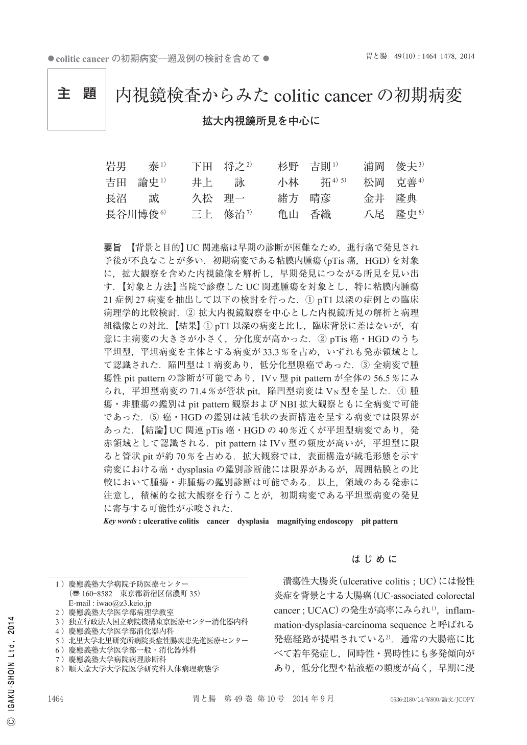
要旨 【背景と目的】UC関連癌は早期の診断が困難なため,進行癌で発見され予後が不良なことが多い.初期病変である粘膜内腫瘍(pTis癌,HGD)を対象に,拡大観察を含めた内視鏡像を解析し,早期発見につながる所見を見い出す.【対象と方法】当院で診療したUC関連腫瘍を対象とし,特に粘膜内腫瘍21症例27病変を抽出して以下の検討を行った.(1)pT1以深の症例との臨床病理学的比較検討.(2)拡大内視鏡観察を中心とした内視鏡所見の解析と病理組織像との対比.【結果】(1)pT1以深の病変と比し,臨床背景に差はないが,有意に主病変の大きさが小さく,分化度が高かった.(2)pTis癌・HGDのうち平坦型,平坦病変を主体とする病変が33.3%を占め,いずれも発赤領域として認識された.陥凹型は1病変あり,低分化型腺癌であった.(3)全病変で腫瘍性pit patternの診断が可能であり,IVV型pit patternが全体の56.5%にみられ,平坦型病変の71.4%が管状pit,陥凹型病変はVN型を呈した.(4)腫瘍・非腫瘍の鑑別はpit pattern観察およびNBI拡大観察ともに全病変で可能であった.(5)癌・HGDの鑑別は絨毛状の表面構造を呈する病変では限界があった.【結論】UC関連pTis癌・HGDの40%近くが平坦型病変であり,発赤領域として認識される.pit patternはIVV型の頻度が高いが,平坦型に限ると管状pitが約70%を占める.拡大観察では,表面構造が絨毛形態を示す病変における癌・dysplasiaの鑑別診断能には限界があるが,周囲粘膜との比較において腫瘍・非腫瘍の鑑別診断は可能である.以上,領域のある発赤に注意し,積極的な拡大観察を行うことが,初期病変である平坦型病変の発見に寄与する可能性が示唆された.
Background and Aims : The prognosis of UCAC(ulcerative colitis-associated cancer)is generally poor because early detection is considerably difficult. We analyzed magnifying endoscopic findings of IEN(intraepithelial neoplasia)in early UCAC to determine the endoscopic characteristics for early detection.
Materials and Methods : Twenty-one IENs from 27 patients were retrospectively reviewed and the clinical and magnifying endoscopic characteristics were compared with invasive neoplasia.
Results : 1)Clinical characteristics did not differ between IENs and invasive neoplasia. 2)All IENs were recognized as reddish areas and 33.3%were flat. 3)All lesions showed neoplastic pit patterns including type IVV(56.5%), round shape(71.4%of flat lesions), and type VN pit patterns(depressed lesions). 4)All IENs were distinguishable from non-neoplastic lesions on the basis of the pit pattern and NBI magnification. 5)It was difficult to distinguish IENs from high-grade dysplasia when they had a villous appearance.
Conclusions : Nearly 40%of IENs were characterized by a flat reddish appearance. The IVV pit pattern was most frequent ; however, the round shape pit pattern was observed in 70%of flat lesions. Magnifying endoscopy was useful for distinguishing neoplastic from non-neoplastic lesions, but not when IENs showed a villous appearance. Our results suggest that magnifying endoscopic observation of reddish areas may improve the early detection of flat UCAC and dysplasia.

Copyright © 2014, Igaku-Shoin Ltd. All rights reserved.


