Japanese
English
- 有料閲覧
- Abstract 文献概要
- 1ページ目 Look Inside
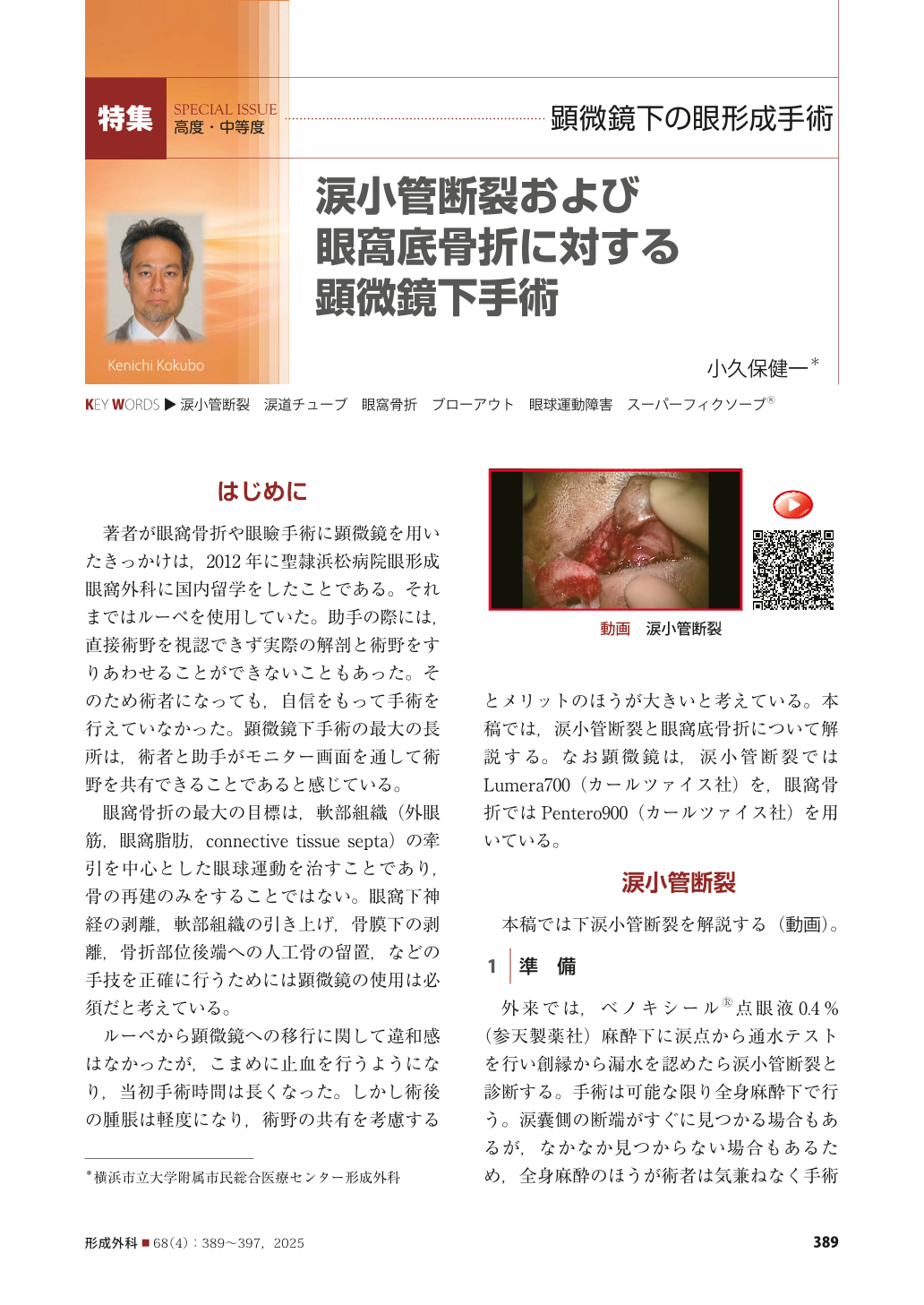
はじめに
著者が眼窩骨折や眼瞼手術に顕微鏡を用いたきっかけは,2012年に聖隷浜松病院眼形成眼窩外科に国内留学をしたことである。それまではルーペを使用していた。助手の際には,直接術野を視認できず実際の解剖と術野をすりあわせることができないこともあった。そのため術者になっても,自信をもって手術を行えていなかった。顕微鏡下手術の最大の長所は,術者と助手がモニター画面を通して術野を共有できることであると感じている。
眼窩骨折の最大の目標は,軟部組織(外眼筋,眼窩脂肪,connective tissue septa)の牽引を中心とした眼球運動を治すことであり,骨の再建のみをすることではない。眼窩下神経の剥離,軟部組織の引き上げ,骨膜下の剥離,骨折部位後端への人工骨の留置,などの手技を正確に行うためには顕微鏡の使用は必須だと考えている。
ルーペから顕微鏡への移行に関して違和感はなかったが,こまめに止血を行うようになり,当初手術時間は長くなった。しかし術後の腫脹は軽度になり,術野の共有を考慮するとメリットのほうが大きいと考えている。本稿では,涙小管断裂と眼窩底骨折について解説する。なお顕微鏡は,涙小管断裂ではLumera700(カールツァイス社)を,眼窩骨折ではPentero900(カールツァイス社)を用いている。
Operating microscopes are used in the eyelid and orbital surgeries conducted in our department. I started using a microscope in 2012 when I studied in Japan at the Department of Ocular Plastic & Orbital Surgery, Seirei Hamamatsu General Hospital. Until then, I sometimes did not have a clear view of the surgical field as an assistant in the operating room. Notably, the use of a microscope allows the surgeon and assistants to share the operative field. This article describes the use of an operating microscope during procedures for lacrimal canalicular lacerations and orbital floor fractures. Serial photographs are included for a clear description.
Copyright© 2025 KOKUSEIDO CO., LTD. All Rights Reserved.