Japanese
English
- 有料閲覧
- Abstract 文献概要
- 1ページ目 Look Inside
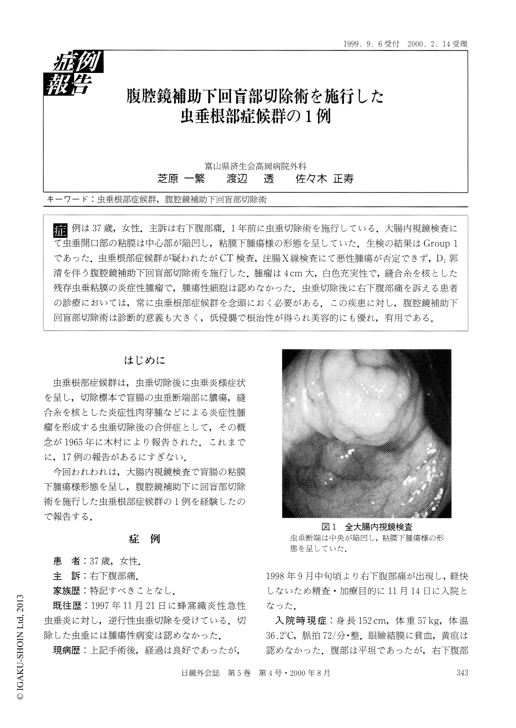
症例は37歳,女性.主訴は右下腹部痛.1年前に虫垂切除術を施行している.大腸内視鏡検査にて虫垂開口部の粘膜は中心部が陥凹し,粘膜下腫瘍様の形態を呈していた.生検の結果はGroup 1であった.虫垂根部症候群が疑われたがCT検査,注腸X線検査にて悪性腫瘍が否定できず,D1郭清を伴う腹腔鏡補助下回盲部切除術を施行した.腫瘤は4cm大,白色充実性で,縫合糸を核とした残存虫垂粘膜の炎症性腫瘤で,腫瘍性細胞は認めなかった.虫垂切除後に右下腹部痛を訴える患者の診療においては,常に虫垂根部症候群を念頭におく必要がある.この疾患に対し,腹腔鏡補助下回盲部切除術は診断的意義も大きく,低侵襲で根治性が得られ美容的にも優れ,有用である.
A 37-year-old female was admitted to our hospital with right lower abdominal pain. She underwent an appendectomy, a year ago. Colonoscopy revealed a stump of appendix, like a submucosal tumor with central depression. The biopsy specimens suggested an inflammatory change of colonic mucosa. Though residual appendicitis was suspected, laparoscopy-assisted ileocecalectomy was performed because of a possibility for malignancy from the results of barium enema and abdominal CT investigation. The resected material showed a solid tumor, 4 cm in size.

Copyright © 2000, JAPAN SOCIETY FOR ENDOSCOPIC SURGERY All rights reserved.


