Japanese
English
- 有料閲覧
- Abstract 文献概要
- 1ページ目 Look Inside
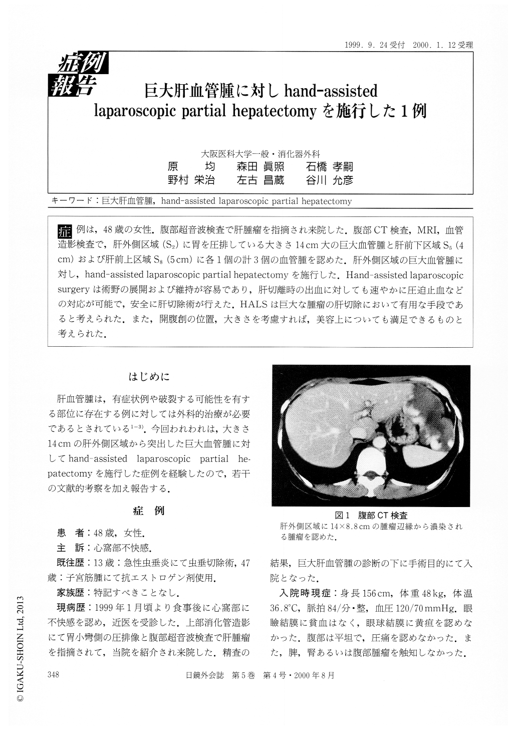
症例は,48歳の女性.腹部超音波検査で肝腫瘤を指摘され来院した.腹部CT検査,MRI,血管造影検査で,肝外側区域(S2)に胃を圧排している大きさ14cm大の巨大血管腫と肝前下区域S5(4cm)および肝前上区域S8(5cm)に各1個の計3個の血管腫を認めた.肝外側区域の巨大血管腫に対し,hand-assisted laparoscopic partial hepatectomyを施行した.Hand-assisted laparoscopicsurgeryは術野の展開および維持が容易であり,肝切離時の出血に対しても速やかに圧迫止血などの対応が可能で,安全に肝切除術が行えた.HALSは巨大な腫瘤の肝切除において有用な手段であると考えられた.また,開腹創の位置,大きさを考慮すれば,美容上についても満足できるものと考えられた.
A 48-year old female was referred to our hospital because of a liver tumor which had been identified by an abdominal ultrasonography. Abdominal computed tomography (CT), magnetic resonance imaging (MRI), and celiac angiography revealed a huge hepatic hemangioma, measuring 14 cm in size and compress-ing the stomach, in the left lateral segment (S2) and two other hemangiomas, measuring 4 cm and 5 cm in size, respectively, in the right anterior inferior segment (S5), and in the right anterior superior segment (S8). The huge hemangioma in the left lateral segment was removed under a hand-assisted laparoscopic surgery.

Copyright © 2000, JAPAN SOCIETY FOR ENDOSCOPIC SURGERY All rights reserved.


