Japanese
English
- 有料閲覧
- Abstract 文献概要
- 1ページ目 Look Inside
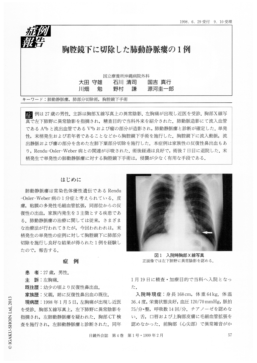
症例は27歳の男性.主訴は胸部X線写真上の異常陰影.左胸痛が出現し近医を受診.胸部X線写真で左下肺野に異常陰影を指摘され,精査目的で当科外来を紹介された.肺動脈造影にて流入血管であるA8bと流出血管であるV8bおよび瘤の部分が造影され,肺動静脈瘻と診断が確定した.単発性,末梢発生および若年者であることなどから胸腔鏡下手術を施行した.胸腔鏡下に流入動脈,流出静脈および瘻の部分を含めた左肺下葉部分切除を施行した.本症例は家族性の反復性鼻出血もあり,Rendu-Osler-Weber病との関連が示唆された.術後経過は良好で,術後7日目に退院した.末梢発生で単発性の肺動静脈瘻に対する胸腔鏡下手術は,侵襲が少なく有用な手段である.
A 27-year-old male was admitted to our hospital with an abnormal shadow on chest X-ray. Chest X-ray showed a nodular lesion in the left lower lung. Chest CT and pulmonary angiography (PAG) revealed a nodular lesion which was diagnosed as pulmonary arteriovenous fistula. Feeding artery was A8b and drainage vein was V8b in the left S8. We performed a partial resection of the left lower lobe (S8), including the feeding artery and the drainage vein. Postoperative course was uneventful. The results indicated that thoracoscopic resection was useful in the surgical treatment of simple pulmonary arteriovenous fistula of the peripheral lung.

Copyright © 1999, JAPAN SOCIETY FOR ENDOSCOPIC SURGERY All rights reserved.


