Japanese
English
原著
気道内魚骨異物症例の検討とX線診断に関する実験的考察
CLINICAL AND EXPERIMENTAL STUDIES ON RADIO-GRAPHIC DIAGNOSIS OF FISH-BONE IN AIRWAY
木村 訓子
1
,
朴沢 二郎
1
,
斎藤 久樹
1
,
袴田 勝
1
,
川畑 武裕
2
,
坂本 哲夫
3
,
樽沢 清
3
Noriko Kimura
1
1弘前大学医学部耳鼻咽喉科学教室
2大館市立病院耳鼻咽喉科
3弘前大学診療放射線部
pp.677-681
発行日 1980年9月20日
Published Date 1980/9/20
DOI https://doi.org/10.11477/mf.1492209132
- 有料閲覧
- Abstract 文献概要
- 1ページ目 Look Inside
Ⅰ.はじめに
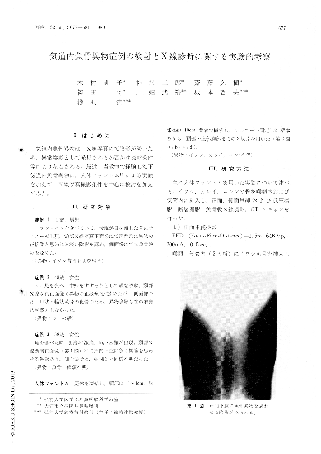
気道内魚骨異物は,X線写真にて陰影が淡いため,異常陰影として発見されるか否かは撮影条件等により左右される。最近,当教室で経験した下気道内魚骨異物に,人体ファントム1)による実験を加えて,X線写真撮影条件を中心に検討を加えてみた。
The following findings were obtained from 3 cases of fish-bone in airway and from some experiments using a human phantom.
1) When the long axis of fish-bone stayed coincidentally with the direction of X-ray beam, it could be found in a frontal view of routine X-ray film or a lateral view of low voltage X-ray film.
2) When the beam of X-ray crossed the long axis of fish-bone, it could be detected only by X-ray tomography.
3) CT-scan in this study was not so useful in comparison with X-ray tomography.

Copyright © 1980, Igaku-Shoin Ltd. All rights reserved.


