Japanese
English
- 有料閲覧
- Abstract 文献概要
- 1ページ目 Look Inside
- 参考文献 Reference
Ⅰ.はじめに
唾液腺あるいはその導管に異物が迷入することは稀であるため1),このような症例に遭遇したときには診断が遅延し,治療に難渋することが多い。今回われわれは術前の画像診断が有効で,早期に摘出し得た顎下腺内魚骨異物症例を経験したので報告する。
□Abstract
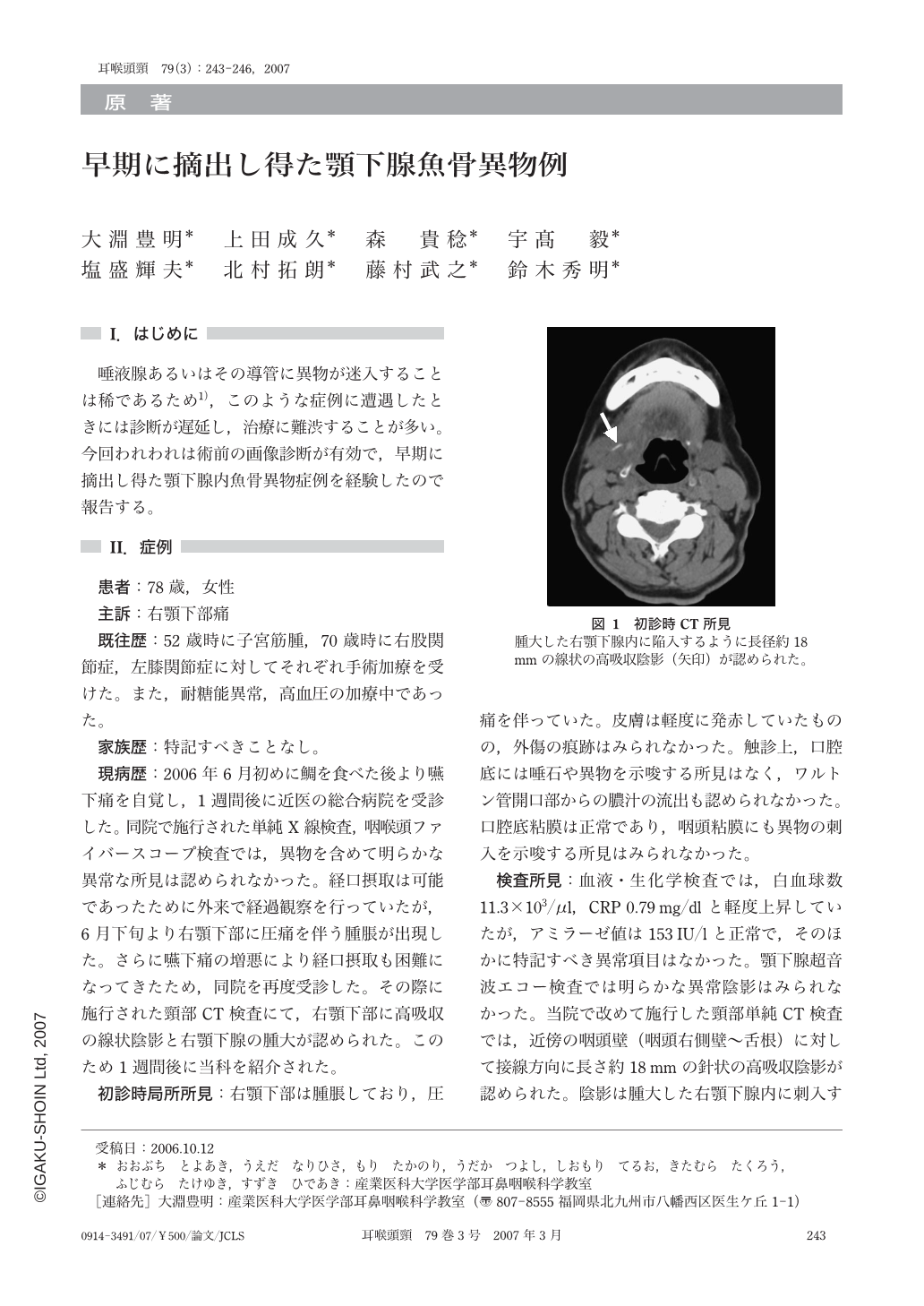
A 78-year-old woman was complaining of painful swallowing after she had eaten a fish. No foreign body was found upon physical examinations, pharyngolaryngeal fiberscopy, or X-rays. However, 2 weeks later, she showed a right-sided submandibular painful swelling, and referred to our department 27 days after onset. CT and CT sialography revealed that a needle-like high-density material staying in the submandibular gland but was separated from the Wharton's duct. A migratory fish bone was suspected, and the patient underwent resection of the gland 33 days after onset. An 18mm-long fish bone was found in the surgical specimen. Her clinical course was uneventful, and she was discharged on the 7th postoperative day. We should be aware that a foreign body penetrating oral or pharyngeal mucosa may migrate to a distant site of the neck.
Copyright © 2007, Igaku-Shoin Ltd. All rights reserved.