Japanese
English
- 有料閲覧
- Abstract 文献概要
- 1ページ目 Look Inside
I.はじめに
通常,髄膜腫は孤発性に発生するが,Neurofibroma—tosis(以下NFと略す)に合併して家族性に発生する例が知られている.
われわれは,NFの徴候を欠く母娘に生じた前頭円蓋部の富血管性5)(angiomatous)髄膜腫を経験したので,文献的考察を含めて報告する.
The convexity angiomatous meningiomas that occur-red in a mother and a daughter without any evidence of neurofibromatosis (NF) were reported.
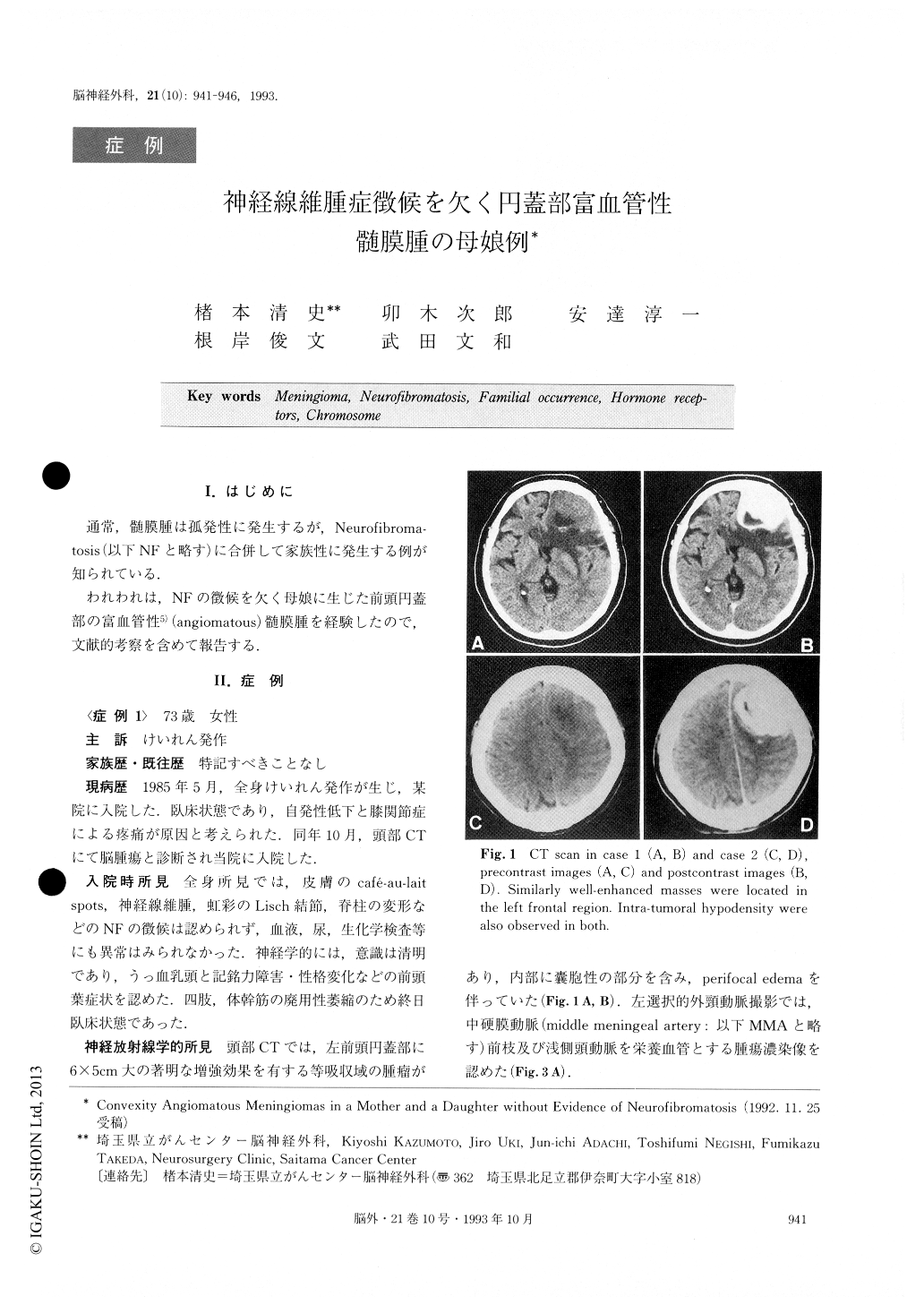
The 73-year-old mother was admitted to our clinic be-cause of an episode of generalized convulsion and a five-month history of gait disturbance. She had the signs/ symptoms of intracranial hypertension and frontal lobe dysfunctions. Computed tomography (CT) revealed a left frontal enhanced mass with a small intratumoral cyst and a remarkable perifocal edema. Angiography showed tumor stain fed from the external carotid artery. Frontal craniotomy was performed and a dark red tumor was totally resected. The nodular-surfaced tumor had adhered loosely to the dura mater. The coarse vascular meshwork and an intratumoral cyst were observed on the cut surface. When the patient was discharged she was able to leave the hospital on foot, but she died of acute pancreatitis in the local hospital. Histological ex-amination of the tumor showed rich vasculatures with focally whorl-formatecl cells. Most tumor cells had intracy-toplasmic microcysts. The pathological diagnosis, WHO's classification, 1991, was angiomatous menin-gioma.
The patient's 41-year-old daughter was admitted due to an episode of fainting. All laboratory data were within normal limits, including the normal karyotype of the peripheral blood leukocytes. Papilloeclemata were the only signs of neurological deficit. A CT scan and magne-tic resonance images showed a left frontal convexity mass and angiography displayed the tumor stains from the middle meningeal artery. The convexity meningioma similar to her mother's was totally removed. The histolo-gical diagnosis was angiomatous menigioma, again. She has been free of recurrence for the 25 months since the surgery.
It is well known that meningiomas may occur in the same family members in cases of type 2 of NF. These meningiomas with NF are usually the fibroblastic type, and are reported to be 13% of all NF patients with brain tumors analyzed in Japan, 1977.
Familial meningiomas in parent and children without evidence of NF, however, have been reported in only 26 patients out of 11 families including the present case. Wefound three clinical preponderances among these cases : the female predominancy, onset earlier in the second-generation patients than in the first, and the histopatho-logical similarity. We speculate that these features may indicate some genetic factors relating to the origin of the tumors. Coincidental loss of genes on chromosome 22 was demonstrated in over half of the meningiomas. We need further study to find out genetic changes of somatic cells in familial cases without any evidence of NF, whichmay lead to our being able to predict the occurrence of meningiomas.
The present cases had similarity with respect to clinic-al courses, tumor locations, radiological and histopatho-logical findings. Angiomatous meningiomas in a parent and a child with no evidence of NF have never been pre-viously reported in the literature.
Copyright © 1993, Igaku-Shoin Ltd. All rights reserved.