Japanese
English
- 有料閲覧
- Abstract 文献概要
- 1ページ目 Look Inside
I.はじめに
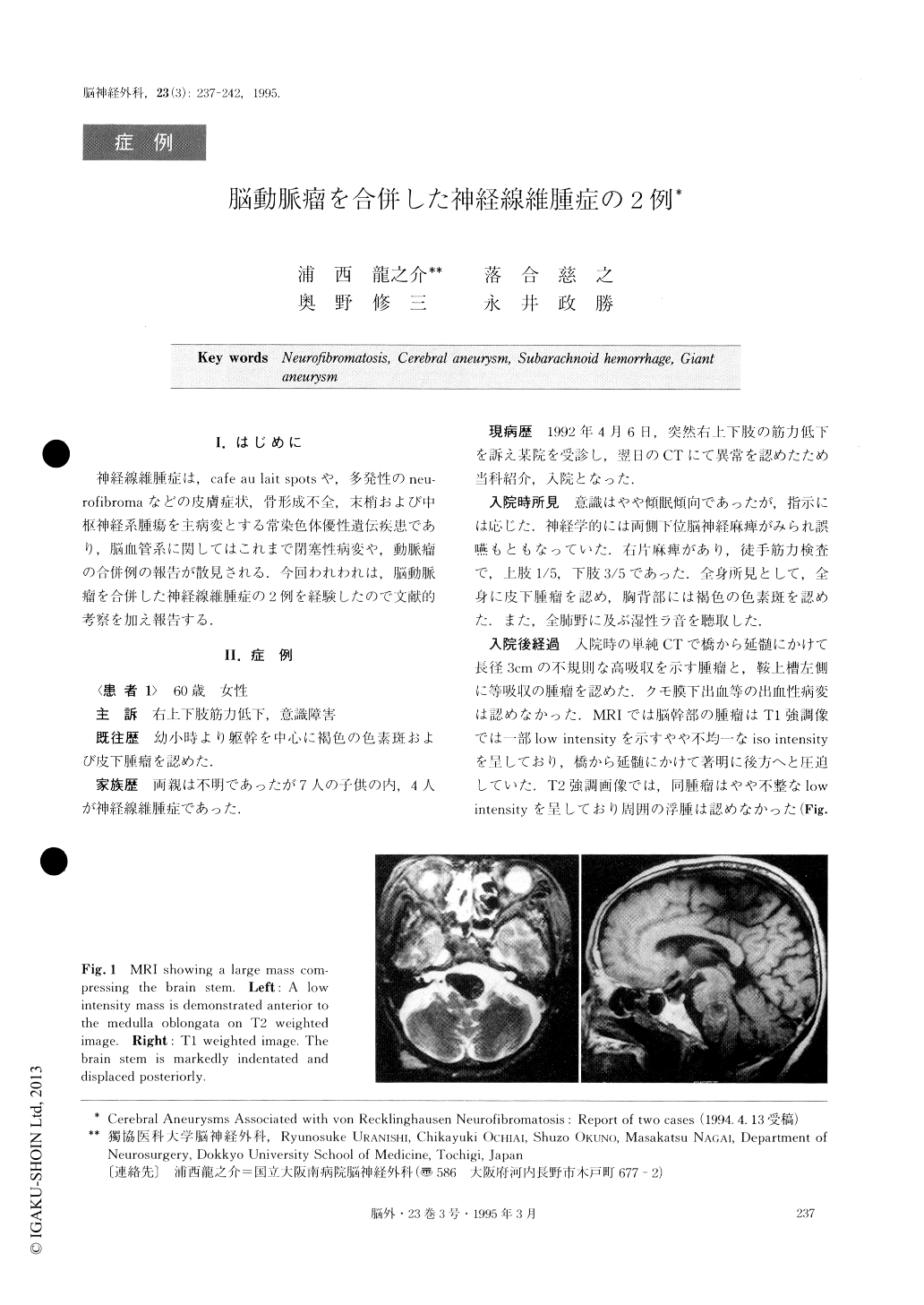
神経線維腫症は,cafe au lait spotsや,多発性のneu—rofibromaなどの皮膚症状,骨形成不全,末梢および中枢神経系腫瘍を主病変とする常染色体優性遺伝疾患であり,脳血管系に関してはこれまで閉塞性病変や,動脈瘤の合併例の報告が散見される.今回われわれは,脳動脈瘤を合併した神経線維腫症の2例を経験したので文献的考察を加え報告する.
The authors reported two cases of aneurysms associ-ated with von Recklinghausen's neurofibromatosis. The first case, a 60-year-old woman was referred to our hos-pital because of disturbance of consciousness and right hemiparesis of acute onset. On admission, she was almost alert but developed right hemiparesis and lower cranial nerve palsy. Computed tomographic (CT) scan-ning and magnetic resonance imaging (MRI) demon-strated a huge mass compressing the brain stem and another mass lesion in the suprasellar cistern. Cerebral angiography disclosed left vertebral and left internal carotid artery giant aneurysms. Since she suffered re-spiratory distress because of aspiration pneumonia, con-servative therapy was carried out. However, she de-veloped cardiac arrest suddenly and died fourteen days after admission. On autopsy, it was shown that the left sided medulla oblongata had necrosis due to compres-sion by the giant aneurysm and that the hemorrhagic infarction of the left cerebellar hemisphere was caused by a thrombus from the giant aneurysm of the left ver-tebral artery.
The second case, a 40-year-old woman presented a disturbance of consciousness. A CT scanning demon-strated subarachnoid hemorrhage with a thick hemato-ma in the left sylvian fissure. An aneurysm at the junc-tion of the right internal carotid artery and the pos-terior communicating artery was found, while the left middle cerebral artery was shown to be normal on cere-bral angiography. Additionally an arteriovenous fistula of the left vertebral artery was found. Although no aneurysm was seen in the territory of the left internal carotid artery, left sided craniotomy was performed based on the CT findings. No aneurysm was found in-traoperatively. The patient died one week later because of rebleeding.
In the second case, a pathological examination of the affected vessels revealed a characteristic finding, viz ex-cept for proliferation of schwann cells, including intimal hyperplasia, thinning of media and fragmentation of elastic lamina similar to that mentioned in previous re-ports of pathological findings in the renal artery in cases of von Recklinghausen's neurofibromatosis. We think that aneurysmal formation in cases of neurofibro-matosis may be caused by the fragility of the arterial wall because of the mesodermal abnormality.
Copyright © 1995, Igaku-Shoin Ltd. All rights reserved.