Japanese
English
- 有料閲覧
- Abstract 文献概要
- 1ページ目 Look Inside
I.はじめに
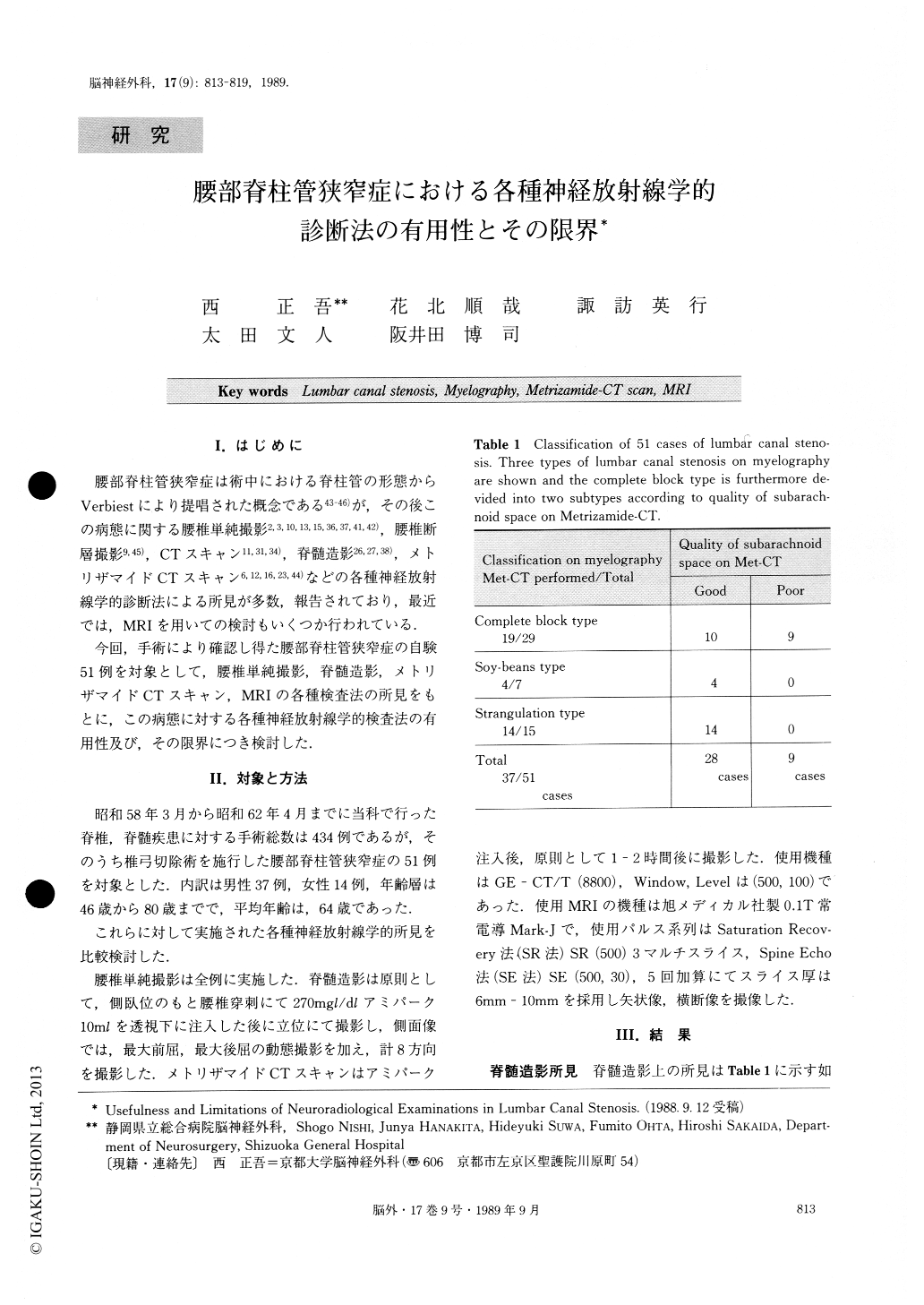
腰部脊柱管狭窄症は術中における脊柱管の形態からVerbiestにより提唱された概念である43-46)が,その後この病態に関する腰椎単純撮影2,3,10,13,15,36,37,41,42),腰椎断層撮影9,45),CTスキャン11,31,34),脊髄造影26,27,38),メトリザマイドCTスキャン6,12,16,23,44)などの各種神経放射線学的診断法による所見が多数,報告されており,最近では,MRIを用いての検討もいくつか行われている,
今回,手術により確認し得た腰部脊柱管狭窄症の自験51例を対象として,腰椎単純撮影,脊髄造影,メトリザマイドCTスキャン,MRIの各種検査法の所見をもとに,この病態に対する各種神経放射線学的検査法の有用性及び,その限界につき検討した.
Since 1983, we have performed 434 spinal surgery operations. Among them are included 51 cases of lum-bar canal stenosis. For these 51 cases, we performed several neuroradiological examinations, such as lumbar plain X-ray, myelography, metrizamide-CT scan (Met-CT) and magnetic resonance imaging (MRI).
We discussed and examined the usefulness and limitations of such neuroradiological methods for di-agnosis of lumbar canal stenosis.
On myelography, these 51 patients were divided into three types; a complete block type with 29 patients, soy-beans type with 7 patients and strangulation type with 15 patients.

Copyright © 1989, Igaku-Shoin Ltd. All rights reserved.


