Japanese
English
症例
血管撮影上著明な腫瘍陰影を呈した傍鞍部脊索腫の1例
Hypervascular Chordoma: Report of a case
平井 収
1,2
,
菊池 晴彦
1
,
山下 純宏
1
Osamu HIRAI
1,2
,
Haruhiko KIKUCHI
1
,
Junkoh YAMASHITA
1
1京都大学脳神経外科
2現籍 浜松労災病院脳神経外科
1Department of Neurosurgery, Faculty of Medicine, Kyoto University
2Department of Neurosurgery, Hamamatsu Rosai Hospital
キーワード:
Chordoma
,
Angiography
,
Magnetic resonance imaging
,
Hypervascularity
,
Radiation therapy
Keyword:
Chordoma
,
Angiography
,
Magnetic resonance imaging
,
Hypervascularity
,
Radiation therapy
pp.757-761
発行日 1988年5月10日
Published Date 1988/5/10
DOI https://doi.org/10.11477/mf.1436202632
- 有料閲覧
- Abstract 文献概要
- 1ページ目 Look Inside
I.はじめに
MRIやX線CTの普及とともに脳腫瘍の診断にも長足の進歩がみられるが,傍鞍部においてはさまざまな病変が発生するために診断に苦慮する場合も多い1,12).
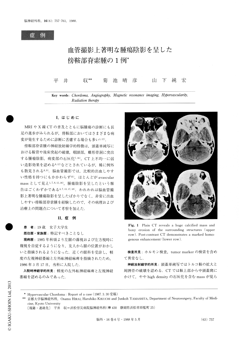
傍鞍部脊索腫の神経放射線学的特徴は,頭蓋単純写における鞍背や後床突起の破壊,咽頭部,蝶形骨洞に突出する腫瘤陰影,病変部の石灰化7,16),CT上不均一に弱い造影効果を認める6,12)などとされているが,稀に例外も散見される8,9).脳血管撮影では,比較的出血しやすい性格を持つにもかかわらず15),ほとんどがavascularmassとして見え1,7,8,12,16),腫瘍陰影を呈したという報告はごくわずかである4,7,13,17,18).われわれは脳血管撮影上著明な腫瘍陰影を呈したばかりでなく,非常に出血しやすい傍鞍部脊索腫を経験したので,その病理および治療上の問題点について考察を加えた.
The authors report a rare case of sellar chordoma with a marked vascularity documented on cerebral angiogram. Its possible pathogenesis and some ther-apeutic problems were briefly discussed. Only a few cases of chordoma with such a positive tumor stain have been reported previously.

Copyright © 1988, Igaku-Shoin Ltd. All rights reserved.


