Japanese
English
出血
―症例―MRIで適切に診断し得た特発性脊髄内出血の1例
Spontaneous Hematomyelia Properly Diagnosed with Magnetic Resonance Imaging
平井 収
1,2
,
菊池 晴彦
1
,
石川 正恒
1
,
米川 泰弘
1,3
Osamu HIRAI
1,2
,
Haruhiko KIKUCHI
1
,
Masatsune ISHIKAWA
1
,
Yasuhiro YONEKAWA
1,3
1京都大学脳神経外科
2現籍 浜松労災病院脳神経外科
3現籍 国立循環器病センター脳神経外科
1Department of Neurosurgery, Faculty of Medicine Kyoto University
3Department of Neurosurgery, National Cardio-Vascular Center
キーワード:
Spontaneous hematomyelia
,
Cervical cord
,
Magnetic resonance imaging
,
Anterior approach
Keyword:
Spontaneous hematomyelia
,
Cervical cord
,
Magnetic resonance imaging
,
Anterior approach
pp.535-538
発行日 1988年5月1日
Published Date 1988/5/1
DOI https://doi.org/10.11477/mf.1436202597
- 有料閲覧
PPV購入案内
1,320円 (1,200円+税10%) こちらは、535ページから538ページの文献です。
PPV(ペイ・パー・ビュー)とは、論文(記事)単位で購入・閲覧ができるサービスです。
購入には医書.jp本会員登録が必要です。
会員登録完了後に改めて上記「購入サイトへ」を選択してください。
または「購入サイトへ」選択後に「購入ログイン」、「新規会員登録」の順に進んでください。
会員登録について詳しくはをご確認ください。
購入には医書.jp本会員登録が必要です。
会員登録完了後に改めて上記「購入サイトへ」を選択してください。
または「購入サイトへ」選択後に「購入ログイン」、「新規会員登録」の順に進んでください。
会員登録について詳しくはをご確認ください。
- Abstract 文献概要
- 1ページ目 Look Inside
I.はじめに
特発性脊髄内出血は通常,脊髄血管腫 5,8,10,14),脊髄空洞症12),抗凝固療法3,13),凝固異常16)などに合併することが多く,原因不明で真に特発性脊髄内出血といえるものは極めて稀である2,9,15).われわれはMRIで経過を追うことにより,初めて適切に診断治療し得た特発性脊髄内出血の1例を経験したので報告する.
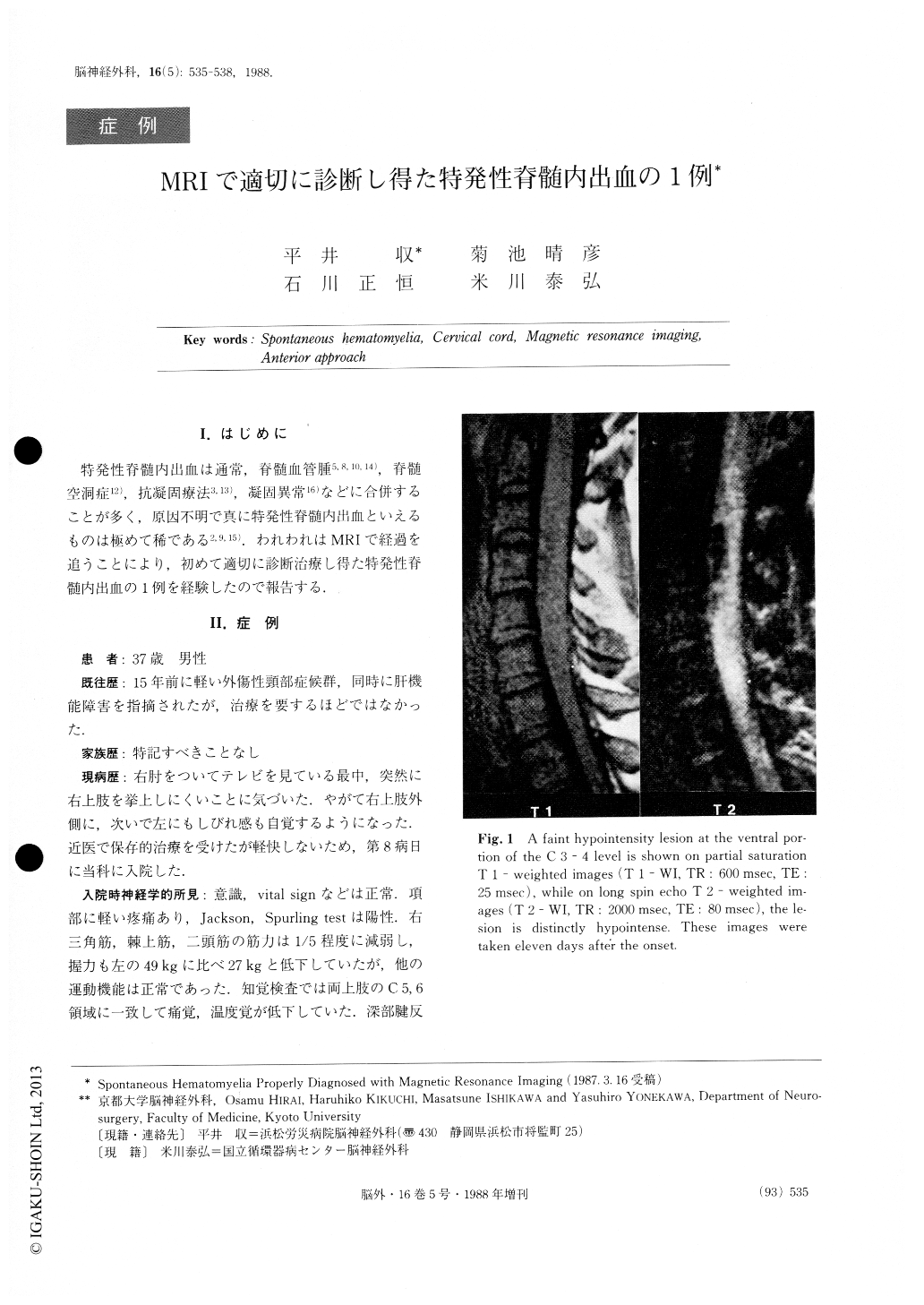
A case of spontaneous hematomylia with symptoms localized to the C 5 - 6 level is presented. Myelography and metrizamide CT scan failed to demonstrate the le-sion, while magnetic resonance imaging clearly revealed a small intramedullary mass in the ventral portion of C 3 - 4. Subsequent intensity changes over four weeks indicated the lesion to be hematoma. The patient was successfully treated through an anterior approach.
Copyright © 1988, Igaku-Shoin Ltd. All rights reserved.