Japanese
English
- 有料閲覧
- Abstract 文献概要
- 1ページ目 Look Inside
I.はじめに
1945年Scheinker21)がsubependymal glia由来と考えられる2例の第4脳室腫瘍を報告し,その組織学的特徴よりsubependymomaという新しいentityを提唱した.本腫瘍は中年以降の年代で,剖検時偶然発見されることが多く,そのほとんどは第4脳室にみられる17).その他に,側脳室,透明中隔,モンロー孔,中脳水道,脊髄にも発生し,種々の神経症候を呈し,外科的治療をうけたものも少なくない1-4,6-8,10-12,15,18,21-23,25,26).
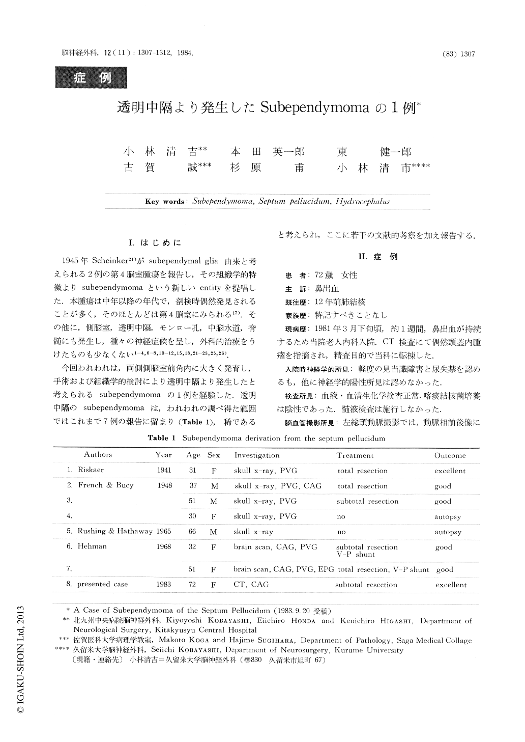
今回われわれは,両側側脳室前角内に大きく発育し,手術および組織学的検討により透明中隔より発生したと考えられるsubependymomaの1例を経験した.透明中隔のsubependymomaは,われわれの調べ得た範囲ではこれまで7例の報告に留まり(Table 1),稀であると考えられ,ここに若干の文献的考察を加え報告する.
A 72-year-old-female was admitted because of slight disorientation and urinary incontinence.
CT scan on admission revealed isodensity mass in the portion of the septum pellucidum and dilated lateral ventricle with patcy enhancement. A patient underwent the right frontal osteplastic craniotomy and subtotal removal of the tumor. Microscopical examination of the removed tumor tissue revealed a typical finding of subepndymoma.
In the previous literature subepndymoma of the septum pellucidum has been reported only seven cases.

Copyright © 1984, Igaku-Shoin Ltd. All rights reserved.


