Japanese
English
- 有料閲覧
- Abstract 文献概要
- 1ページ目 Look Inside
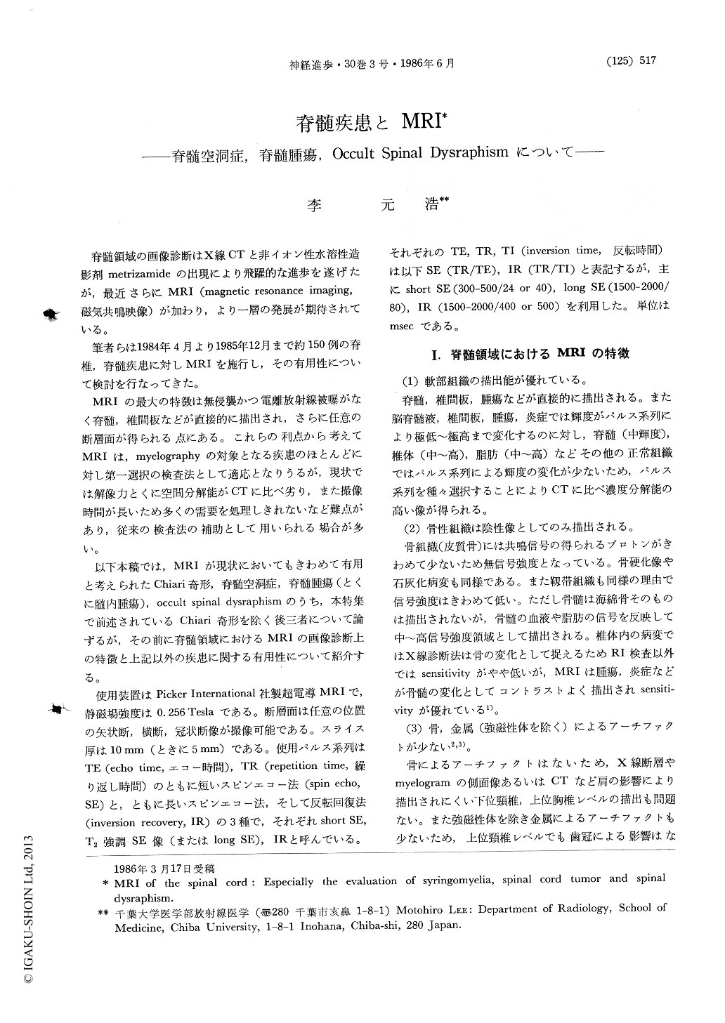
脊髄領域の画像診断はX線CTと非イオン性水溶性造影剤metrizamideの出現により飛躍的な進歩を遂げたが,最近さらにMRI(magnetic resonance imaging,磁気共鳴映像)が加わり,より一層の発展が期待されている。
筆者らは1984年4月より1985年12月まで約150例の脊椎,脊髄疾患に対しMRIを施行し,その有用性について検討を行なってきた。
152 cases with spinal disorders were examined by magnetic resonance imaging (MRI). MRI was very useful in the evaluation of syringomyelia, spinal cord tumor and spinal dysraphism.
Syringomyelia: In all eight cases of this lesion, which were proved by surgery, the cavities were evident on MRI and were obviously distinguished from intramedullary spinal cord tumor. Sagittal images were convenient for seeing the extent of the cavity and the exisistence of Chiari malformation. The signal intensity of the cavity was lower than that of spinal cord on spin echo image with short TR and TE in seven cases.

Copyright © 1986, Igaku-Shoin Ltd. All rights reserved.


