Japanese
English
- 有料閲覧
- Abstract 文献概要
- 1ページ目 Look Inside
はじめに
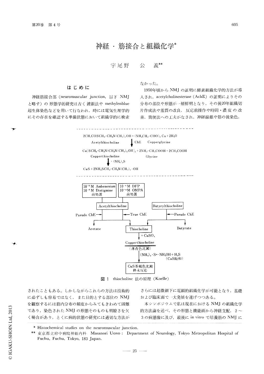
神経筋接合部(neuromuscular junction,以下NMJと略す)の形態学的研究は古く鍍銀法やmethylenblue超生体染色などを用いて行なわれ,時には電気生理学的にその存在を確認する準備状態において組織学的に検索されたこともある。しかしながらこれらの方法は技術的に必ずしも容易ではなく,また目的とする部位のNMJを観察するには筋内分布の頻度からみてもきわめて困難であり,染色されたNMJの形態そのものも明瞭さを欠く場合があり,とくに病的状態の研究には適切な方法がなかった。
1950年頃からNMJの証明に酵素組織化学的方法が導入され,acetylcholinesterase(AchE)の証明によりその分布の部位や形態が一層鮮明となり,その後20年組織切片作成法や基質の改良,反応前操作や時間・濃度の改善,簡便法への工夫がなされ,神経線維や筋の後染色,さらには超微細下に電顕的組織化学が可能となり,基礎および臨床面で一大発展を遂げつつある。
On the studies of structure, function and growing process of neuromuscular junction (NMJ),I have approached, this time, mainly by using methods of histochemistry, electronmicroscopic histochemistry and also histochemistry for the cultured tissue, and got the following results.
1. As for the histochemical method on NMJ, I can get most specific and stable results by applying the modified method which uses acetyl-thiocholine (Koelle-Tsuji or Karnovsky-Tsuji method) for the time being.

Copyright © 1976, Igaku-Shoin Ltd. All rights reserved.


