Japanese
English
- 有料閲覧
- Abstract 文献概要
- 1ページ目 Look Inside
I.緒言
神経筋疾患の研究は近年急速な発展をみている。その理由として,筋生検がさかんに行なわれるようになつたことと10 26 61 94),生検あるいは剖検筋に対し従来の形態学的・生化学的検索のみならず,組織化学的・電子顕微鏡的・免疫化学的に広角度から検討が加えられるようになつたことなどがあげられる25)33)59)61)74)38)90)91)97)。
したがつてこれら疾患の成因的分類もかなり詳細に整理されてきたが,一方研究方法の進歩とともに従来の考え方では説明し得ない新たな疾患も数多く報告されている29)70)71)72)。
The activities of phosphorylase (PhR) (Take-uchi & Kuriaki stain, 1956), succinic dehydro-genase (SDH) (Nachlas et al stain, 1957) and cholinesterase (ChE) (Koelle stain, 1951. Karnov-sky stain, 1964) were assessed by histochemical techniques in biopsied and autopsied muscles of 94 patients with various neuromuscular diseases.
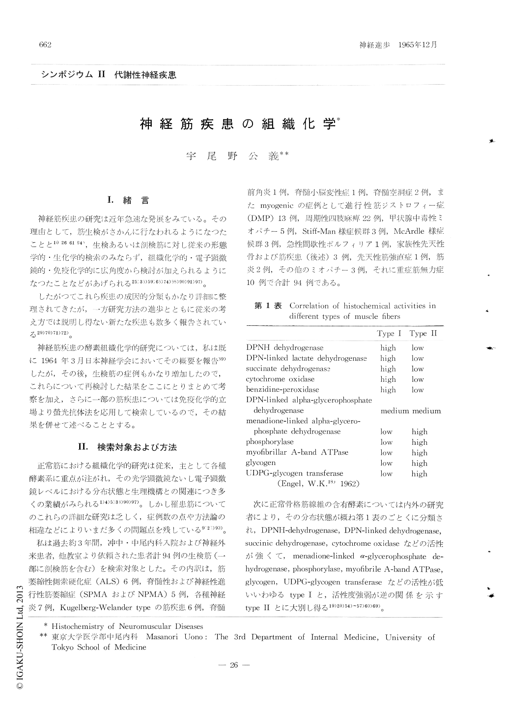
Amyotrophic lateral sclerosis (ALS) 6 Spinal and Neural progressive muscular atrophy (SPM, NPM) 5 Poliomyelitis anterior 1 Neuritis 7 Kugelberg-Welander type muscular atrophy 6 Spinocerebellar degeneration 1 Syringomyelia 2 Progressive muscular dystrophy 13 Periodic paralysis 22 Thyrotoxic myopathy 5 Stiff-Man-like syndrome 3 McArc-lie-like syndrome 3 Acute intermittent porphyria 1 Congenital familiar osteomyopathy 3 Myotonia congenita 1 Myositis 2 Miscellaneous myopathies 3 Myasthenia gravis 10 Total 94
The results obtained were as follows: 1) In neurogenic atrophic muscle fibers (i.e.ALS, SPM, NPM, Poliomyelitis, Kugelberg-Welancler type muscular atrophy), reductin of SDH activity was evidently seen. There was little change of PhR activity and of ChE activity in motor end-plates until atrophy was severe.
2) It is well known the structur and function of external eye muscles in ALS is kept better than those of the other skeletal muscles. In my results, PhR, SDH and ChE were much more active and no deformity of endplates was seen in external eye muscles with ALS, too.
3) In dystrophic muscle, there was little change in SDH activity until atrophy was severe, but PhR became much generally more inactive, howe-ver some enlarged and small fibers remained still strong in PhR. The relationship between the density of PhR and SDH activities and size of dystrophic muscle fivers shown by Dubowitz & Pearse was not immediately evident.
4) In periodic paralysis, PhR and SDH activi-ties in muscle fibers and ChE activity in motor end-plates were normal and no morphological findings in end-plates was seen.
5) In myasthenic muscle, PhR and SDH activi-ties were normal but ChE activity in nerve endings and motor end-plates was low and various morphological changes (atrophy, swelling, segmen-taion, elaboration, elongation etc.) were seen.
It would be of great interest to classify and diagnose the neuromuscular diseases by using enzymo-histochemical technique as well as mor-phological and biochemical studies.
On the other hand, immunological studies on several neuromuscular diseases as autoimmune disease were assessed by fluorescence antibody technique (Kawamura et al method, 1963).
Fluorescein labelled serum r-globulin in 5 out of 6 cases with myasthenia gravis combined with homo- and heterozygous (i.e. other human or dogs) skeletal muscle fibers respectively, and not with liver, kidney, lung etc.
Same results were also obtained in fluorescein labelled serum r-globulin in rats with experimental allergic myositis.
Copyright © 1965, Igaku-Shoin Ltd. All rights reserved.