Japanese
English
- 有料閲覧
- Abstract 文献概要
- 1ページ目 Look Inside
はじめに
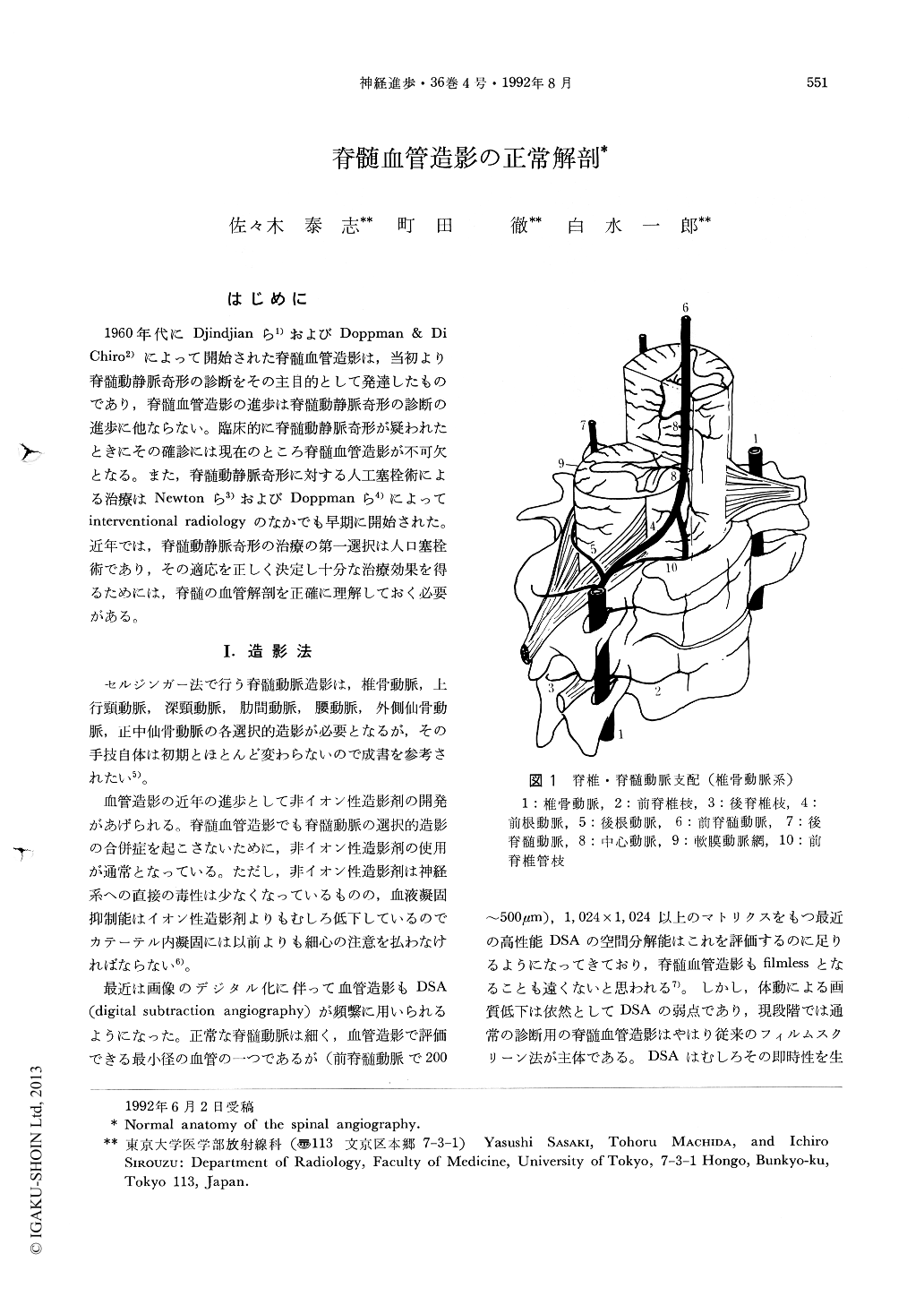
1960年代にDjindjianら1)およびDopPman & Di Chiro2)によって開始された脊髄血管造影は,当初より脊髄動静脈奇形の診断をその主目的として発達したものであり,脊髄血管造影の進歩は脊髄動静脈奇形の診断の進歩に他ならない。臨床的に脊髄動静脈奇形が疑われたときにその確診には現在のところ脊髄血管造影が不可欠となる。また,脊髄動静脈奇形に対する人工塞栓術による治療はNewtonら3)およびDoppmanら4)によってinterventional radiologyのなかでも早期に開始された。近年では,脊髄動静脈奇形の治療の第一選択は人口塞栓術であり,その適応を正しく決定し十分な治療効果を得るためには,脊髄の血管解剖を正確に理解しておく必要がある。
The authors review recent advancements of the spinal angiography. Nonionic contrast material has been used to decrease the risk of complication on spinal angiography. Digital subtraction angiography has been applied to spinal angiography in recent years, but its main role is for the interventional procedures.
The authors also review the essential radiological anatomy of the spinal vasculature. The spinal arterial system is developed from 31 segmental arteries, and these are composed of the vertebral, ascending cervical, deep cervical, intercostal, lumbar, lateral sacral, median sacral arteries. Anterior and posterior radiculomedullary arteries arise from some of these segmental arteries, and the greatest anterior radiculomedullary artery in the thoracolumbar spine is called the artery of Adamkiewicz. The spinal arteries are the arterial axes along the spinal cord. The anterior spinal artery is located on the midline of the spinal canal fed by the anterior radiculomedullay arteries, and the both posterior spinal arteries are slightly off the midline fed by ipsilateral posterior radiculomedullary arteries. The former gives rise to the central arteries that penetrate the cord through the anterior median sulcus feeding 2/3 of the cord.
Copyright © 1992, Igaku-Shoin Ltd. All rights reserved.