Japanese
English
- 有料閲覧
- Abstract 文献概要
- 1ページ目 Look Inside
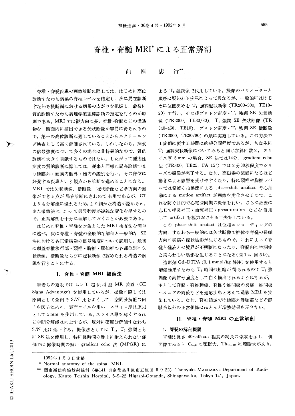
脊椎・脊髄疾患の画像診断に際しては,はじめに高位診断すなわち病巣の脊椎レベルを確定し,次に局在診断すなわち横断面における病巣の広がりを把握し,最後に質的診断すなわち病理学的組織診断の推定を行うのが原則である。MRIでは縦方向に長い脊椎・脊髄などの構造物を一断面内に描出できる矢状断像が容易に得られるので,第一の高位診断に適していることからスクリーニング検査として高く評価されている。しかしながら,病変の信号強度について多くの場合は非特異的なので,質的診断に大きく貢献するものではない。したがって腫瘤性病変の質的診断に際しては,従来と同様に局在診断つまり硬膜外・硬膜内髄外・髄内の鑑別を行い,その部位に好発する疾患という観点から診断を進めることになる。MRIでは矢状断像,横断像,冠状断像など多方向の撮像ができる点が局在診断にきわめて有用であるが,CTよりも分解能に優れるため,より細かな構造が認められ,また撮像法によって信号強度が複雑な変化を呈するので,正常解剖を十分に理解しておくことが必要である。
はじめに脊椎・脊髄を対象としたMRI検査法を簡単に述べ,次に脊椎・脊髄の全般的な解剖と一般的なSE法における各正常構造の信号強度について説明し,最後に頭蓋脊椎移行部・頸椎・胸椎・腰仙椎の各部位別に矢状断像,横断像ならびに冠状断像で認められる構造の解説を行うことにする。
Magnetic resonance imaging (MRI) is the most valuable imaging modality in the diagnosis of spinal cord lesions. For utilizing this diagnostic modality effectively, it is very important to understand the normal anatomy as well as basic science aspects of the modality.
The purpose of this paper is to illustrate the MR appearance of the spine and its contents and to provide guidelines for interpreting spinal MRI. It contains the method of obtaining the optimal spinal MR images, the general remarks of the normal spinal MR anatomy including the signal inten-sity of the cord, nerve roots, intervertebral discs and CSF in the dural sac, and detailed atlas sections. The MR images in this paper were acquired with GE Signa Advantage (1.5 T) system using spin-echo pulse sequences.

Copyright © 1992, Igaku-Shoin Ltd. All rights reserved.


