Japanese
English
- 有料閲覧
- Abstract 文献概要
- 1ページ目 Look Inside
- 参考文献 Reference
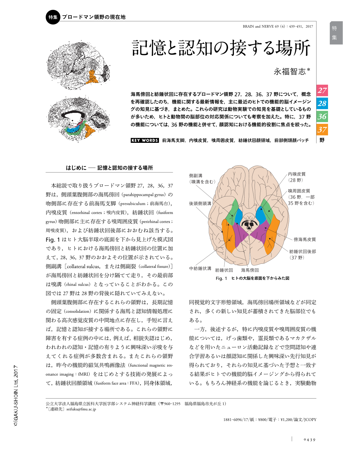
海馬傍回と紡錘状回に存在するブロードマン領野27,28,36,37野について,概念を再確認したのち,機能に関する最新情報を,主に最近のヒトでの機能的脳イメージングの知見に基づき,まとめた。これらの研究は動物実験での知見を基礎としているものが多いため,ヒトと動物間の脳部位の対応関係についても考察を加えた。特に,37野の機能については,36野の機能と併せて,顔認知における機能的役割に焦点を絞った。
Abstract
First, Brodmann areas 27, 28, 36 and 37, were anatomically defined in the beginning of this review. These areas exist in the parahippocampal or fusiform gyrus of the ventral temporal lobe in humans. Subsequently, the current understanding of their functions was summarized on the basis of recent findings mainly through human functional neuroimaging studies and animal studies. Rodent studies have shown the existence of neuronal activities for representing space, such as those involving head-direction cells or grid cells, in areas 27 (the parasubicular cortex) and 28 (the ventral entorhinal cortex). Recent human neuroimaging studies have provided support for the idea that grid cells may also exist in the human entorhinal cortex. Many previous animal studies have shown that area 36 (the lateral perirhinal cortex) is crucial for various types of associative learning. Earlier human neuroimaging studies have also indicated that faces, bodies and visual word forms are represented in different regions of area 37 in the posterior fusiform gyrus. Recent neuroimaging studies in humans have shown substantial functional differentiation between face-related regions in areas 37 and 36, which is similar to that seen in macaque monkeys, as shown through their face patches. This implies the crucial involvement of both areas in face processing.

Copyright © 2017, Igaku-Shoin Ltd. All rights reserved.


