Japanese
English
- 有料閲覧
- Abstract 文献概要
- 1ページ目 Look Inside
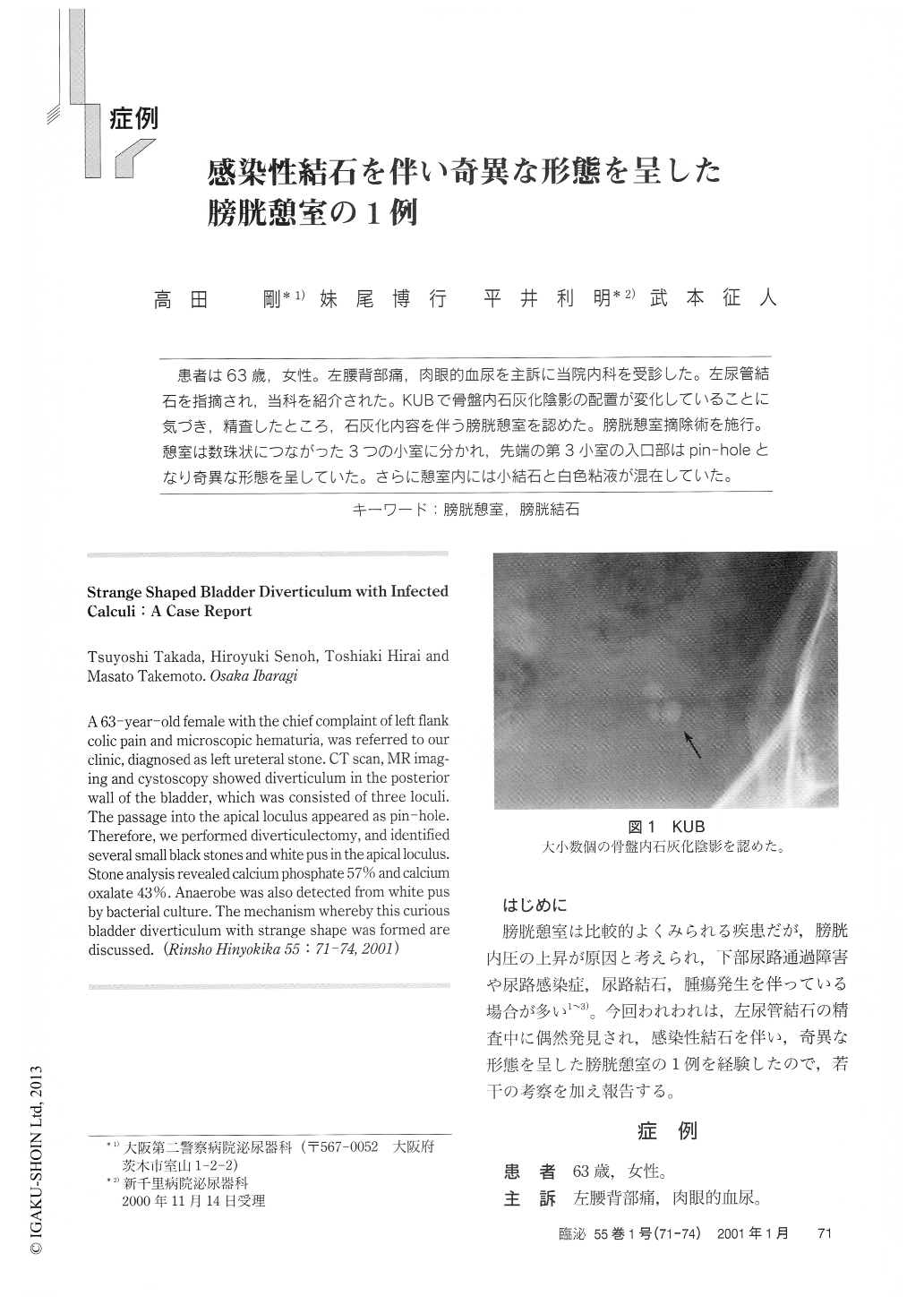
患者は63歳,女性。左腰背部痛,肉眼的血尿を主訴に当院内科を受診した。左尿管結石を指摘され,当科を紹介された。KUBで骨盤内石灰化陰影の配置が変化していることに気づき,精査したところ,石灰化内容を伴う膀胱憩室を認めた。膀胱憩室摘除術を施行。憩室は数珠状につながった3つの小室に分かれ,先端の第3小室の入口部はpin-holeとなり奇異な形態を呈していた。さらに憩室内には小結石と白色粘液が混在していた。
A 63-year-old female with the chief complaint of left flank colic pain and microscopic hematuria, was referred to our clinic, diagnosed as left ureteral stone. CT scan, MR imag-ing and cystoscopy showed diverticulum in the posterior wall of the bladder, which was consisted of three loculi.The passage into the apical loculus appeared as pin-hole.Therefore, we performed diverticulectomy, and identified several small black stones and white pus in the apical loculus.Stone analysis revealed calcium phosphate 57% and calcium oxalate 43%. Anaerobe was also detected from white pus by bacterial culture.

Copyright © 2001, Igaku-Shoin Ltd. All rights reserved.


