Japanese
English
- 有料閲覧
- Abstract 文献概要
- 1ページ目 Look Inside
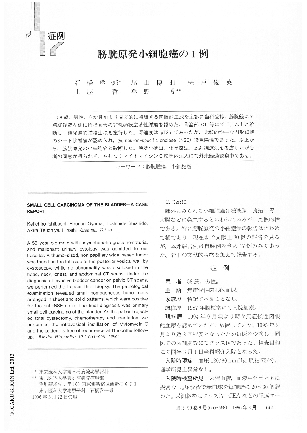
58歳,男性。6か月前より間欠的に持続する肉眼的血尿を主訴に当科受診。膀胱鏡にて膀胱後壁左側に拇指頭大の非乳頭状広基性腫瘍を認めた。骨盤部CT等にてT2以上と診断し,経尿道的腫瘍生検を施行した。深達度はpT3aであったが,比較的均一な円形細胞のシート状増殖が認められ,抗neuron-specific enolase(NSE)染色陽性であった。以上から,膀胱原発の小細胞癌と診断した。膀胱全摘出,化学療法,放射線療法を考慮したが患者の同意が得られず,やむなくマイトマイシンC膀胱内注入にて外来経過観察中である。
A 58-year-old male with asymptomatic gross hematuria, and malignant urinary cytology was admitted to our hospital. A thumb-sized, non papillary wide-based tumor was found on the left side of the posterior vesical wall by cystoscopy, while no abnormality was disclosed in the head, neck, chest, and abdominal CT scans. Under the diagnosis of invasive bladder cancer on pelvic CT scans, we performed the transurethral biopsy. The pathological examination revealed small homogeneous tumor cells arranged in sheet and solid patterns, which were positive for the anti-NSE stain.

Copyright © 1996, Igaku-Shoin Ltd. All rights reserved.


