Japanese
English
- 有料閲覧
- Abstract 文献概要
- 1ページ目 Look Inside
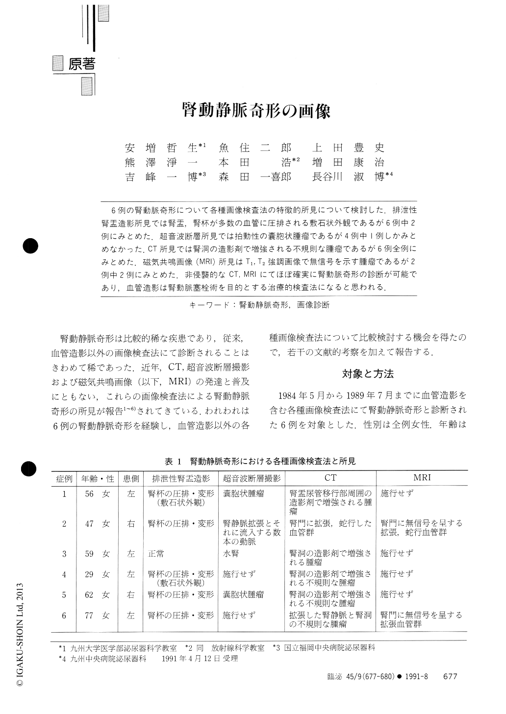
6例の腎動脈奇形について各種画像検査法の特徴的所見について検討した.排泄性腎盂造影所見では腎盂,腎杯が多数の血管に圧排される敷石状外観であるが6例中2例にみとめた.超音波断層所見では拍動性の嚢胞状腫瘤であるが4例中1例しかみとめなかった.CT所見では腎洞の造影剤で増強される不規則な腫瘤であるが6例全例にみとめた.磁気共鳴画像(MRI)所見はT1,T2強調画像で無信号を示す腫瘤であるが2例中2例にみとめた.非侵襲的なCT,MRIにてほぼ確実に腎動脈奇形の診断が可能であり,血管造影は腎動脈塞栓術を目的とする治療的検査法になると思われる.
Diagnostic value of various imaging techniques for renal arteriovenous malformation (AVM) was evaluated in 6 cases. Excretory pyelograhy showed typical cobble stone appearance in 2 of the 6 patients. Ultrasonography revealed a cystic mass associated with visible pulsations in central complex in one of the 4 patients. Computed tomography (CT) demonstrated an irregular soft density mass which was enhaned by contrast medium in renal sinus in all of the 6 patients. Magnetic resonance imaging (MRI) was performed on 2 patients and revealed an irregular mass as a region of low intensity on both T1 and T2 weighted images in these patients.

Copyright © 1991, Igaku-Shoin Ltd. All rights reserved.


