Japanese
English
- 有料閲覧
- Abstract 文献概要
- 1ページ目 Look Inside
緒言
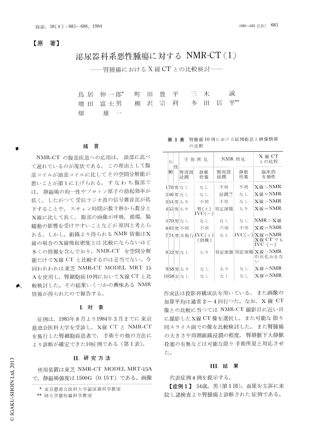
NMR-CTの腹部疾患への応用は,頭部に比べて遅れているのが現状である。この理由として腹部コイルが頭部コイルに比してその空間分解能が悪いことが第1に上げられる。すなわち腹部では,静磁場の均一性やプロトン原子の励起効率が低く,したがつて受信ラジオ波の信号雑音比が低下することや,スキャン時間が数十秒から数分とX線に比して長く,腹部の画像が呼吸,循環,腸蠕動の影響を受けやすいことなどが原因と考えられる。しかし,組織より得られるNMR情報はX線の場合のX線吸収密度とは比較にならないほど多くの情報を含んでおり,NMR-CTを空間分解能だけでX線CTと比較するのは妥当でない。今回われわれは東芝NMR-CT MODEL MRT 15Aを使用し,腎細胞癌10例においてX線CTと比較検討した。その結果いくつかの興味あるNMR情報が得られたので報告する。
NMR imaging was performed in 12 cases (10 cases of renal cell carcinoma and 2 cases of angio. myolipoma) and compared with computed tomography. The NMR imager, Toshiba model MRT 15 A with magnet of 1500 gauss, was used and images were produced in transverse, coronal and sagittal directions with different repetition times, delay times and echo times. The characteristic images associated with renal tumors were demonstrated. The renal contour was usually sharp at normal side and easily distinguished from perinephric fat which appeared white. The cortex and medullary pyramids were distinguished in excellent images.

Copyright © 1984, Igaku-Shoin Ltd. All rights reserved.


