Japanese
English
- 有料閲覧
- Abstract 文献概要
- 1ページ目 Look Inside
緒言
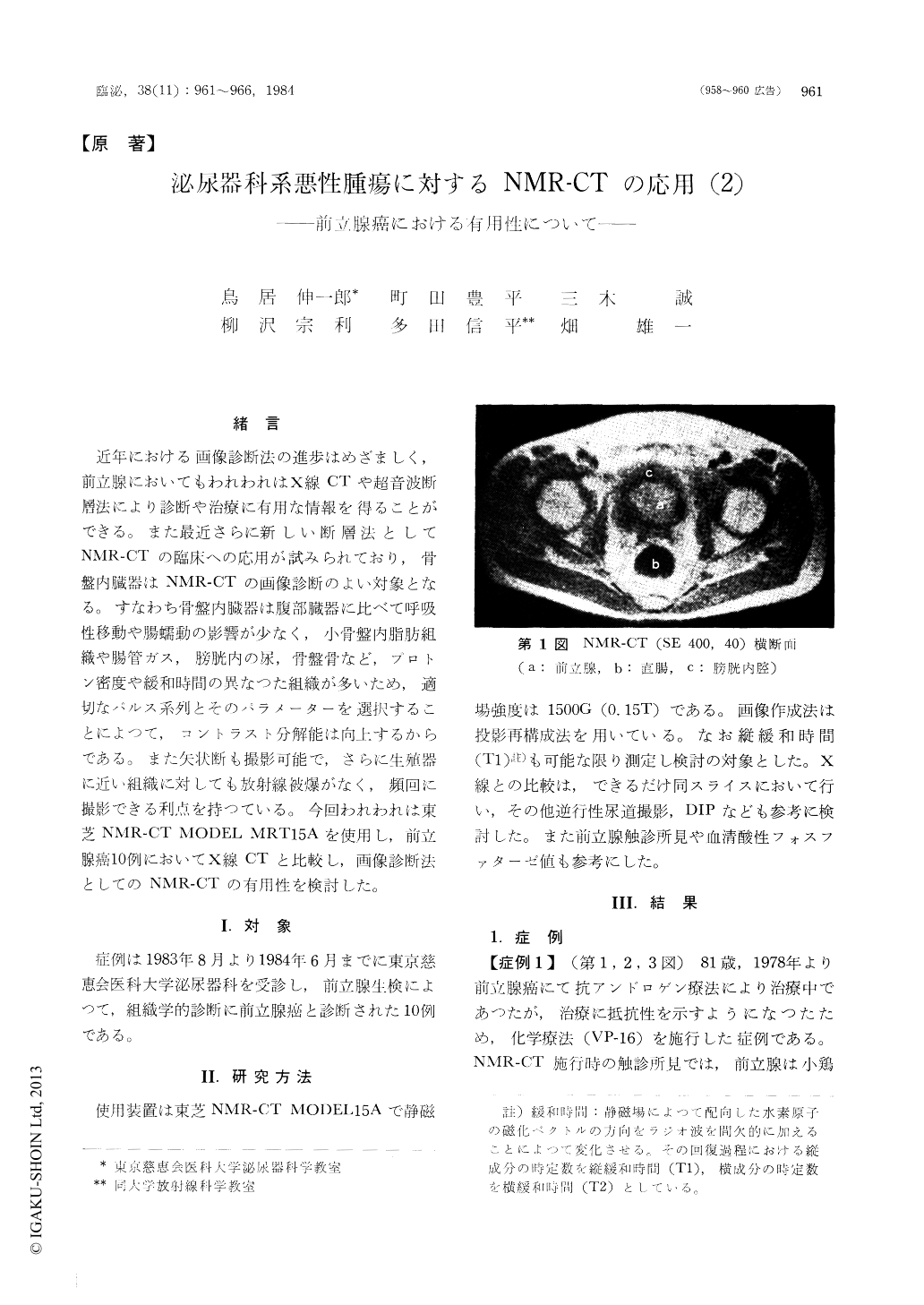
近年における画像診断法の進歩はめざましく,前立腺においてもわれわれはX線CTや超音波断層法により診断や治療に有用な情報を得ることができる。また最近さらに新しい断層法としてNMR-CTの臨床への応用が試みられており,骨盤内臓器はNMR-CTの画像診断のよい対象となる。すなわち骨盤内臓器は腹部臓器に比べて呼吸性移動や腸蠕動の影響が少なく,小骨盤内脂肪組織や腸管ガス,膀胱内の尿,骨盤骨など,プロトン密度や緩和時間の異なつた組織が多いため,適切なパルス系列とそのパラメーターを選択することによつて,コントラスト分解能は向上するからである。また矢状断も撮影可能で,さらに生殖器に近い組織に対しても放射線被爆がなく,頻回に撮影できる利点を持つている。今回われわれは東芝NMR-CT MODEL MRT 15Aを使用し,前立腺癌10例においてX線CTと比較し,画像診断法としてのNMR-CTの有用性を検討した。
Experience and evaluation of NMR imaging of the ten patients with prostatic carcinoma were reported. The NMR imager, Toshiba model MRT 15A with magnet of 1500 gauss, was used. Images were produced in transverse, coronal and sagittal directions with different repetition times, delay times and echo times. T1 value over regions of the prostatic cancer was measured and compared with the value of the benign prostate hypertrophy. In all cases there was high inherent contrast differentiation of prostate gland, pelvic fat, urine in the bladder and gas in the bowel. The characteristic images of the prostatic cancer were demonstrated.

Copyright © 1984, Igaku-Shoin Ltd. All rights reserved.


