Japanese
English
- 有料閲覧
- Abstract 文献概要
- 1ページ目 Look Inside
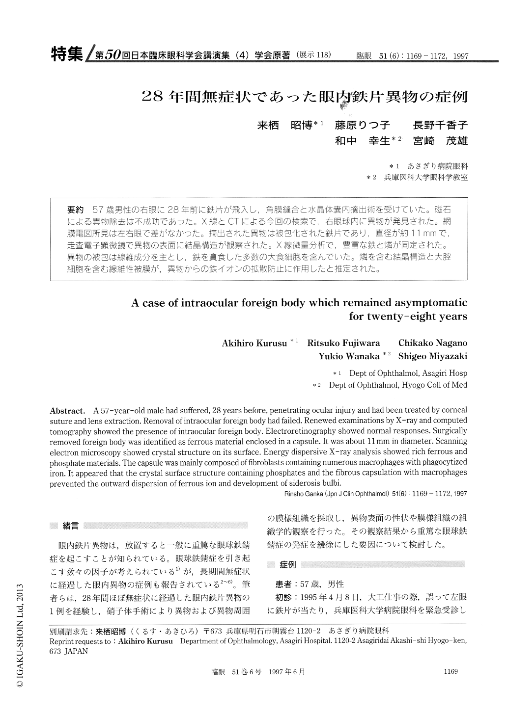
(展示118) 57歳男性の右眼に28年前に鉄片が飛入し,角膜縫合と水晶体嚢内摘出術を受けていた。磁石による異物除去は不成功であった。X線とCTによる今回の検索で,右眼球内に異物が発見された。網膜電図所見は左右眼で差がなかった。摘出された異物は被包化された鉄片であり,直径が約11mmで,走査電子顕微鏡で異物の表面に結晶構造が観察された。X線微量分析で,豊富な鉄と燐が同定された。異物の被包は線維成分を主とし,鉄を貪食した多数の大食細胞を含んでいた。燐を含む結晶構造と大腔細胞を含む線維性被膜が,異物からの鉄イオンの拡散防止に作用したと推定された。
A 57-year-old male had suffered, 28 years before, penetrating ocular injury and had been treated by corneal suture and lens extraction. Removal of intraocular foreign body had failed. Renewed examinations by X-ray and computed tomography showed the presence of intraocular foreign body. Electroretinography showed normal responses. Surgically removed foreign body was identified as ferrous material enclosed in a capsule. It was about 11 mm in diameter. Scanning electron microscopy showed crystal structure on its surface. Energy dispersive X-ray analysis showed rich ferrous and phosphate materials. The capsule was mainly composed of fibroblasts containing numerous macrophages with phagocytized iron. It appeared that the crystal surface structure containing phosphates and the fibrous capsulation with macrophages prevented the outward dispersion of ferrous ion and development of siderosis bulbi.

Copyright © 1997, Igaku-Shoin Ltd. All rights reserved.


